• HOME
  • ABOUT
    • EndoMontreal Clinic
    • Dr. Pierre Pizem
  • CONTACT
  • EN | FR

ENDOMONTREAL

Dr Pizem Advanced Endodontics Procedures

  • Advanced Root Canal Treatment
    • Symptoms & Procedure
    • Why Micro Endodontics?
  • General Dentistry
  • Patients info
    • Frequently asked questions (FAQ)
    • Having a failed root canal treatment?
    • Treatment sequence at our clinic
    • Tooth saving tips
  • For Dentists
    • Refer a patient
    • Clinical cases
You are here: Home / Atypical canal configurations / A Vertucci’s Type III Canal Configuration in Distal Root (Case 410)

A Vertucci’s Type III Canal Configuration in Distal Root (Case 410)

June 24, 2008 By Dr Pierre Pizem Leave a Comment

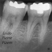
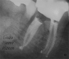
Atypical canal configurations, Type III, Root Canal Treatment Pre-Therapy 410-1Atypical canal configurations, Type III, Root Canal Treatment Post-Therapy 410-1

Vertucci’s Type III canal configuration

One canal leaves the pulp chamber and divide into two in the root; the two then merge to exit as one canal (1-2-1).

According to Vertucci’s study in 1984, the percentage of human permanent teeth with this canal configuration are 5 % for the maxillary second premolar and 0% for mandibular first and second molars. This post operative X Ray shows (once the huge denticle has been removed in coronal third) that the Vertucci’s Type III canal configuration is actually existing in a mandibular first molar.

As stated by Vertucci’s and al.

The pulp canal system is complex, and canals may branch, divide, and rejoin.

Filed Under: Atypical canal configurations, Curved canals, Dental operating microscope (D.O.M.) assisted R.C.T., Extreme endo clinical cases, NEW CASES, Pulp stones (denticles), Type III

Clinical cases categories

  • Atypical canal configurations
    • Radix Entomolaris
    • Taurodontism
    • Type II
    • Type III
    • Type V
    • Type VI (Crossed canals)
    • Very long teeth
    • ‘C’ shaped canal system
  • Curved canals
    • Dilaceration
    • Extremely curved root canals (90 degrees +)
    • C-form canal curvature
    • S-form canal curvature
  • Dental operating microscope (DOM) assisted RCT
    • DOM vs. cracks
    • DOM vs. completely calcified systems
    • DOM vs. partially calcified systems
    • Striving for 2ndMesio vestibular (MB2)
    • Pulp stones (denticles)
  • Dental operating microscope and retreatment
    • Finding previously underseen MB2
    • Orthograde MTA plugs and root repairs
    • Broken instruments removal
    • Casted and machined posts removal
    • Ledges and apical zip
    • N2 (Sargenti Paste) removal
    • Silver points removal
  • Extreme clinical cases
  • Post and core build ups
  • Post endodontic treatment outcomes
    • Regeneration of the periradicular tissues
    • Resorptive defects healings
  • Root canal systems calcifications
  • Surgical removal of tooth structure
    • Tooth sectioning
    • Apicoectomy, curettage and retrofilling

Leave a Reply Cancel reply

Your email address will not be published. Required fields are marked *

© 2019 ENDOMONTREAL · HOME · ABOUT · CONTACT US