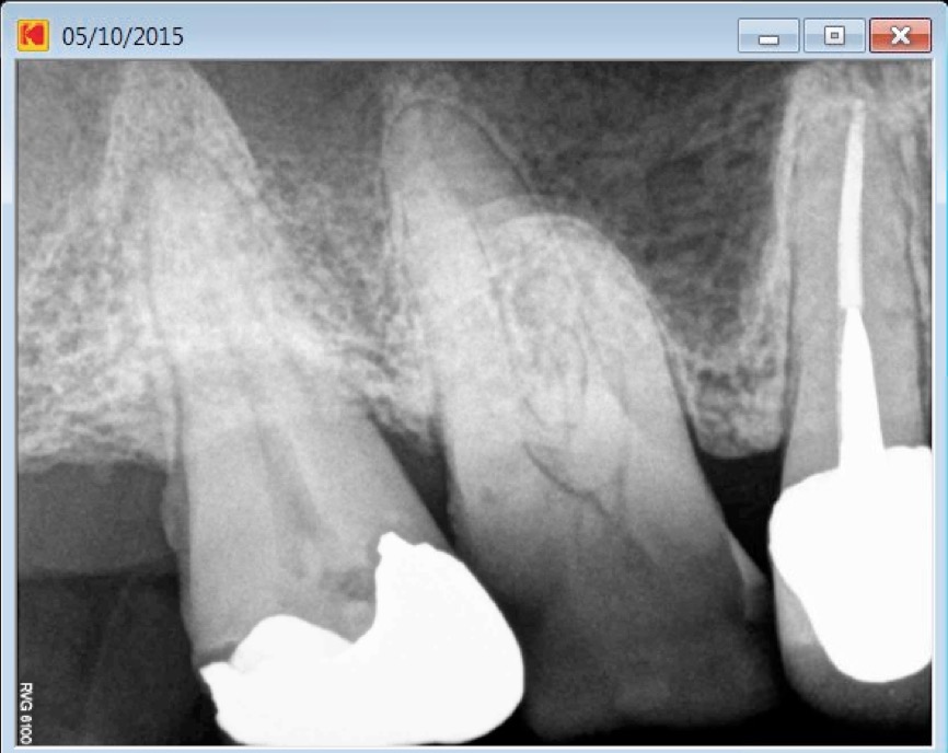
Patient has been referred for root canal treatment on both first and second maxillary molars. Tooth number 16 had necrotic pulp and tooth 17 presented with irreversible pulpitis. Patient was taking both antibiotics and analgesics.
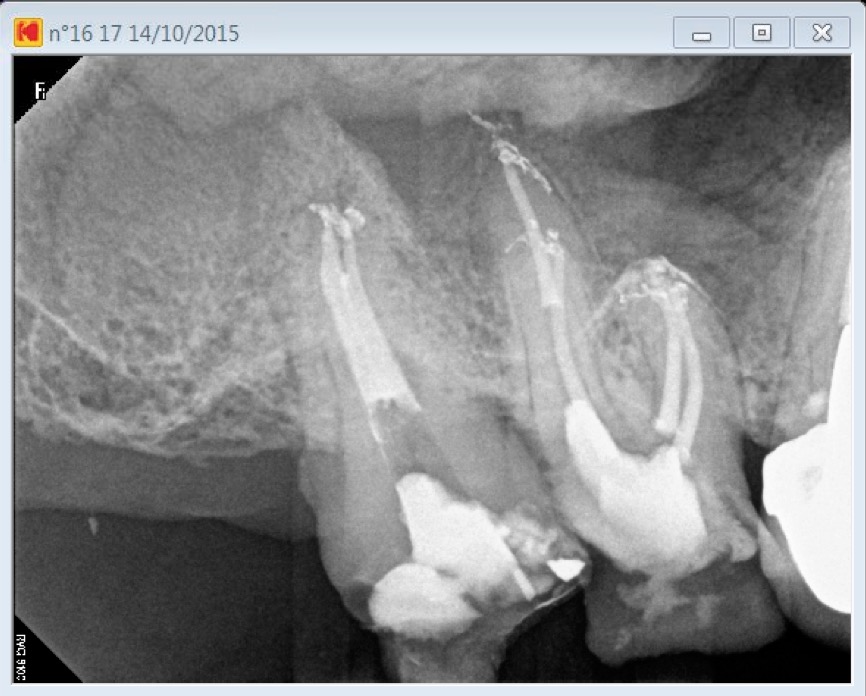
To alleviate patient’s pain, both teeth had to be simultaneously cleaned and shaped during the first setting. A pulpectomy on 17 and a cleaning and shaping of 3 canals of tooth 16 have been performed. An interim calcium hydroxide dressing has been inserted and patient had to come back in order to locate clean and shape the second mesio vestibular. During the second appointment the dental operative microscope allowed us to uncover and treat both an apical split in tooth number 17 and a fourth canal in tooth 16.
Despite the fact that on the pre operative X Ray image tooth number 17 seemed to have a single large canal, it ended up being a Vertucci type 5 root canal configuration (a single canal splitting in two branches short of the apex). As for tooth 16 a second mesio vestibular was expected to be present in its mesio vestibular root, still, dental operative microscope has there too been very useful when trying to locate it.
Access opening has been performed with a combination of a 556 cross cut and a round tungsten carbide bur (Friction Grip). Operative field observation has been enhanced with high magnification and coaxial xenon lamp illumination (Carl Zeiss OPMI PROergo dental operative microscope). Dystrophic calcifications have been removed from pulp chamber with ultrasonic diamond coated tips in order to expose the second mesiovestibular root canal entry location.
Instrumentation has been performed with Mani stainless K files and ProTaper Universal (Dentsply). Chelator: RC Prep. PUI Irrigation: NaOCl 5%. Ultrasonic tip: BUC One from Spartan. P5 (Dentsply) Sealer used: Pulp Canal Sealer. Obturation technique WVC. X Ray Sensors used: CareStream Kodak 6100.
Nice cases
Thank you Dr Ayman!