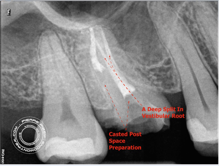
First maxillary molars typically have 4 canal entries and four distinct canals. This molar has only two canal entries and a deep split in its vestibular root (A Vertucci type V root canal configuration). Both canal entries were also embedded in a mass of adherent calcifications. Furthermore, to my dismay, palatal canal last apical instrument size was a F5. In a case such as this one a dental operating microscope has been most helpful when attempting to locate root canal entries as well as when time came to visualize the deep apical split.
Endodontic procedure instrumentation has been performed with Mani stainless K files and ProTaper Universal (Dentsply). Chelator: RC Prep. PUI Irrigation: NaOCl 5%. pulp stones removal with Ultrasonic tip: BUC One from Spartan. P5 (Dentsply) Sealer used: Pulp Canal Sealer. Obturation technique WVC. X Ray Sensors used: CareStream Kodak 6100. Opmi Proergo microscope

Leave a Reply