Case Study Number 186336 A few days ago we were confronted to this three rooted mandibular first molar (Radix Molar or Radix Entomolaris), a
A Vertucci’s Type V Canal Configuration on a Second Maxillary Premolar
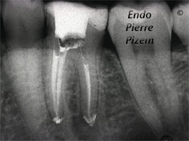
Case Study Number 473515 OPMI PROergo from Carl Zeiss allowed us to clearly see the apical split. Each branch has been shaped, cleaned and filled
Dealing with Dystrophic Calcification in Mesiovestibular Root Canals
Case Study Number 486726 Patient was referred to us with a partial pulpectomy in 3 out of four canals. The case came with a
What’s new on this blog on Friday,11th, 2011
A new case study number: 411716: "The dental operative microscope and the MTA tooth perforation repair. The impossible made possible".
The Dental Operative Microscope and a MTA Tooth Perforation Repair. The Impossible Made Possible.
Case Study Number 461716 Patient came to our office with spontaneous intermittent pain on maxillary right side. He can readily identify the tooth,
Dentistry with Zeiss OPMI PRO Ergo Operative Microscope: Striving to Find a Pathway to the Apex by Bypassing a “Mega Odontolith” in an Anomalous RC System
This is a symptomatic mandibular first premolar (bridge abutment) with a "C" shape root canal system. Most C-shaped canals occurs in mandibular second
Mesial Calcified Canals Entries Location Under High Magnification
An intricate root canal procedure, because this pre operative condition involves dealing with complete canal stenosis caused by dystrophic
Vertucci’s Type V Configuration in Vestibular Root Canal in Maxillary Second Molar
Root canal apical delta could be observed under high magnification during the endodontic procedure. Case Study Number 11917. A Vertucci type V pulp
Two Distinct Right-Angled Root Canals Exits in First Molar Distal Root (Case 455336)
Case Study Number 455336 Irreversible pulpitis, deep carie, deciduous restoration, broken lingual wall. First molar with 4 root canals. Two mesial