• HOME
  • ABOUT
    • EndoMontreal Clinic
    • Dr. Pierre Pizem
  • CONTACT
  • EN | FR

ENDOMONTREAL

Dr Pizem Advanced Endodontics Procedures

  • Advanced Root Canal Treatment
    • Symptoms & Procedure
    • Why Micro Endodontics?
  • General Dentistry
  • Patients info
    • Frequently asked questions (FAQ)
    • Having a failed root canal treatment?
    • Treatment sequence at our clinic
    • Tooth saving tips
  • For Dentists
    • Refer a patient
    • Clinical cases
You are here: Home / Archives for mani stainless endodontic files

A Root Canal Treatment on a Severely Curved Root Canal

August 26, 2018 By Dr Pierre Pizem Leave a Comment

A colleague asked me on my facebook page what has been my root canal treatment approach to overcome that challenging anatomy. https://www.facebook.com/endomontreal/

A) Never push a straight stainless steel file into the canal in order to reach the foramen with it. You must first bind your first instrument tip and scout in order to be able to feel and follow the canal shape or its direction change.

B) In a case such as this one, you will most likely meet more than a point of resistance before reaching the foramen and you will have to carve your way in deeper. Pushing your way in at the point of resistance with your file will definitively create a ledge or else with their disastrous consequences. In order to progress deeper, you have to enlarge the coronal portion with your ss file using the enveloppe of motion technique. Continues this way until the desired length is achieved.

C) Then, once the foramen has been reached, you can enlarge the canal in order to create a glide path up to size 15. Once the glide path has been completed you could use the NiTi shaping file of your choice. Needed state of mind NO pushing but strong resolution.
Endodontic procedure instrumentation has been performed with Mani stainless endodontic K files and ProTaper Universal (Dentsply). Chelator: RC Prep. PUI Irrigation: NaOCl 5%.

Filed Under: Curved canals, Extreme endo clinical cases, Extremely curved root canals (90 degrees +) Tagged With: canal curvature, curvatures of greater than 90 degrees, pulp canal sealer root canal procedure, Root canal treatment, severe curvature, sharply curved canals, Protaper, mani stainless endodontic files

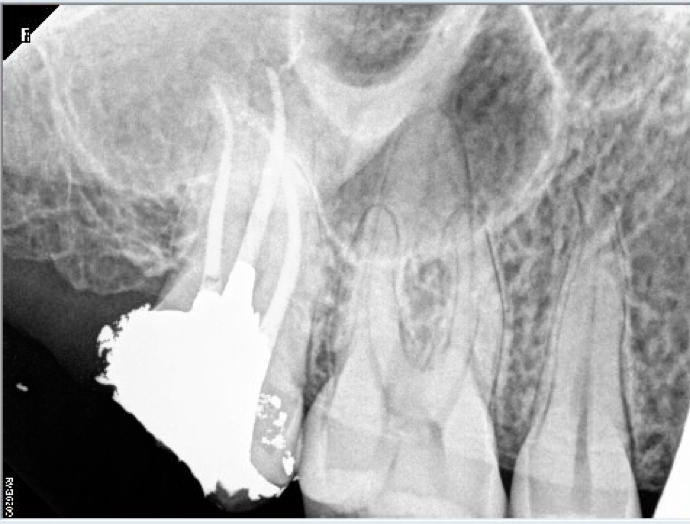
Root canal procedure on a remote maxillary molar in a patient with limited opening of the mouth

April 14, 2017 By Dr Pierre Pizem Leave a Comment


Endodontic procedure instrumentation has been performed with Mani stainless K files and ProTaper Universal (Dentsply). Chelator: RC Prep. PUI Irrigation: NaOCl 5%. Mesio vestibular canal entry located with the help of high magnification and coaxial illumination (Opmi PROergo dental operative microscope from Zeiss) Sealer used: Pulp Canal Sealer. Obturation technique WVC. X Ray Sensors used: CareStream Kodak 6100.

Patient has been advised that the next step would be a crown lengthening and a crown

Filed Under: NEW CASES, Post and core build ups Tagged With: amalgam post, prognosis, Protaper dentsply, root canal perforation, crown lengthening, mani stainless endodontic files, remote area of the mouth, limited opening of the mouth, instrument fracture

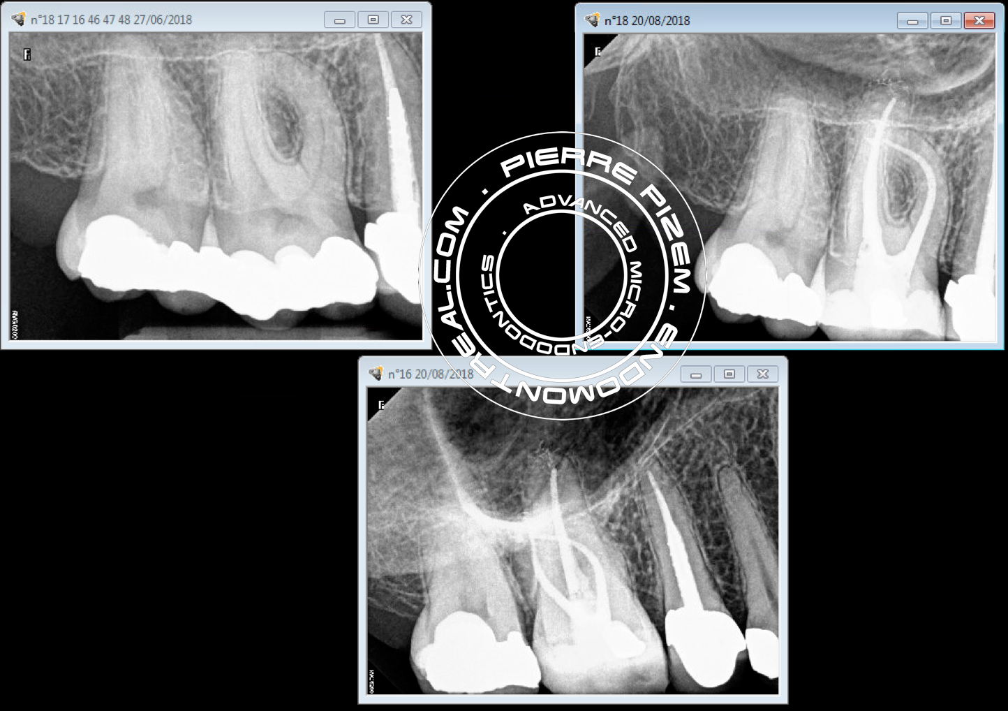
An Extreme Root Canal Treatment Procedure

June 18, 2016 By Dr Pierre Pizem Leave a Comment

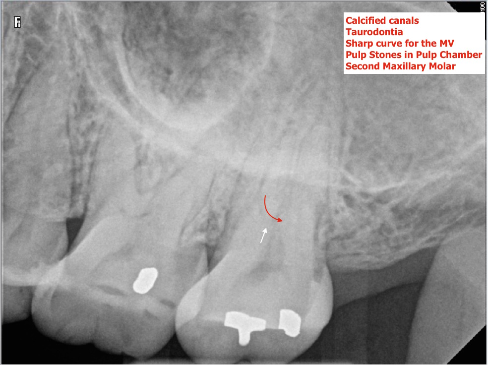
Patient seen on an emergency basis for irreversible pulpitis on second maxillary molar. Pulp stones removal and pulpectomy performed + interim Ca(OH)2 on palatal canal only.
I had to make the patient come back for another setting to complete RCT.
The most difficult part of this procedure has been to obtain patency in a remote mesio vestibular canal as it was located at the bottom of a pulp chamber presenting with taurodontia and also because this canal had a very sharp curve in the first mm of its coronal part. Microscope Opmi ProErgo, Mani K stainless endodontic files, ProTaper from Dentsply.


Root canal treatment procedure on 27 taurodontia and calcified A 2016-06-07  Root canal treatment procedure on 27 taurodontia and calcified B 2016-06-07  Root canal treatment procedure on 27 taurodontia and calcified B 2016-06-07

Filed Under: Atypical canal configurations, Dental operating microscope (D.O.M.) assisted R.C.T., Extreme endo clinical cases, Extremely curved root canals (90 degrees +), NEW CASES, Pulp stones (denticles), Taurodontism, D.O.M. versus partially calcified systems Tagged With: dental operating microscope, opmi proergo, Protaper dentsply, root canal procedure, calcified, mani stainless endodontic files

Root canal procedure on a tooth presenting with a rare anatomical variation

March 29, 2016 By Dr Pierre Pizem Leave a Comment

Tooth 24 long calcified 3 rooted tooth pre operative 2016-03-29 Tooth 24 pre operative fractured cusp 2016-03-29 Tooth 24 long calcified 3 rooted tooth post operative 2016-03-29

Patient had a fractured cusp and has been referred  for a root canal treatment. Indistinct root canal path on pre operative Xray did let us suspect an anatomical variation along with calcified canals. Furthermore, a CareStream Kodak 6100 number 2 sensor had to be positioned vertically in order to get the image of this long tooth to its full length.
Both location and instrumentation of root canal split on buccal root has been performed with the help of an Opmi Proergo microscope magnification.
Both buccal working lengths were 26mm and 28mm for the palatal. Instrumentation has been performed with Mani stainless K files and ProTaper Universal (Dentsply). Chelator: RC Prep. PUI Irrigation: NaOCl 5%. Sealer used: Pulp Canal Sealer. Obturation technique WVC. X Ray Sensors used: CareStream Kodak 6100
During the same setting a root canal space preparation for a casted post and a provisional crown with anchorage have been performed.

Filed Under: Atypical canal configurations, Dental operating microscope (D.O.M.) assisted R.C.T., D.O.M. versus partially calcified systems Tagged With: calcified canals, casted post, crown, Opmi Proergo microscope, pro taper, pulp canal sealer, root canal split, very long tooth, anatomical variation, mani stainless endodontic files, carestream kodak 6100, fractured cusp, buccal root

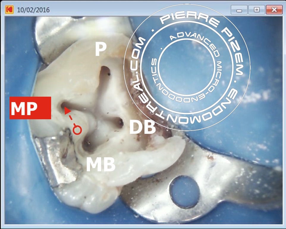
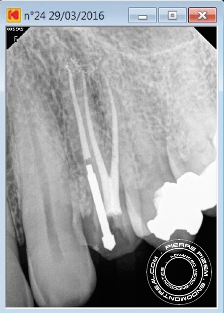
A root canal procedure on a molar presenting with a supernumerary mesio palatal (MP) root

March 10, 2016 By Dr Pierre Pizem 1 Comment

17 second palatal canal pre operative   Root canal procedure on a maxillary molar presenting with a supernumerary palatal root     17 second palatal canal Ca(OH)2 17 second palatal canal post operative

Root canal procedure on molar presenting with an extra mesio palatal (MP) root. A very rare anatomical variation.

This supernumerary root has been noticed on pre operative X ray image and following the grooves on the pulp chamber floor that led to its very large root canal entry made its location  quite straightforward. Extending the access cavity prior to shaping this root canal has been mandatory in order to prevent any apical ledge or endodontic instruments breakage.

Endodontic procedure instrumentation has been performed with Mani stainless K endodontic files and ProTaper Universal (Dentsply). Chelator: RC Prep. PUI Irrigation: Sodium hypochlorite 5%. Sealer used: Pulp Canal Sealer. Obturation technique warm vertical condensation (WVC). X Ray Sensors used: CareStream Kodak 6100


Filed Under: Uncategorized, Atypical canal configurations, Dental operating microscope (D.O.M.) assisted R.C.T., NEW CASES, Radix Entomolaris Tagged With: endodontic procedure, pulp canal sealer, Root canal, root canal procedure, shaping, sodium hypochlorite, supernumerary root, pulp chamber, protaper universal, endodontic instruments, access cavity, mani stainless endodontic files, warm vertical condensation, carestream kodak 6100

© 2019 ENDOMONTREAL · HOME · ABOUT · CONTACT US