• Skip to primary navigation
  • Skip to main content
  • Skip to primary sidebar
  • Skip to footer

  514-667-9541

  REQUEST APPOINTMENT

HOME

ABOUT

DR PIZEM

FRANÇAIS

ENDOMONTREAL

ENDOMONTREAL

Dr Pizem Advanced Endodontics Clinic

  • Advanced Root Canal Treatment
    • Symptoms & Procedure
    • Why Micro Endodontics?
  • General Dentistry
  • Patients info
    • Frequently asked questions (FAQ)
    • Having a failed root canal treatment?
    • Treatment sequence at our clinic
    • Tooth saving tips
  • For Dentists
    • Refer a patient
    • Clinical cases
Contact Us

Dentistry with Zeiss OPMI PRO Ergo Operative Microscope: Striving to Find a Pathway to the Apex by Bypassing a “Mega Odontolith” in an Anomalous RC System

February 2, 2011 By Dr Pierre Pizem Leave a Comment

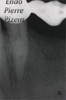
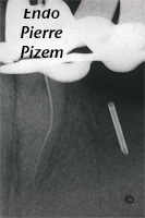
Atypical canal configurations, Type III, Root Canal Treatment Pre-Therapy 156035-1Atypical canal configurations, Type III, Root Canal Treatment Per-Therapy 156035-1Atypical canal configurations, Type III, Root Canal Treatment Per-Therapy 156035-2

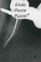
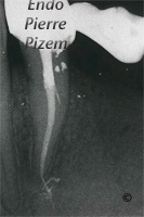
Atypical canal configurations, Type III, Root Canal Treatment Per-Therapy 156035-3Atypical canal configurations, Type III, Root Canal Treatment Per-Therapy 156035-4Atypical canal configurations, Type III, Root Canal Treatment Post-Therapy 156035-1

This is a symptomatic mandibular first premolar (bridge abutment) with a “C” shape root canal system. Most C-shaped canals occurs in mandibular second molars but they have been also reported in the mandibular first molar, the maxillary  first and second molars and the mandibular first premolar. This “C” shape first mandibular premolar root canal system is extremely rare. The only orthograde way to endodontically treat this anomalous root is to bring our first K file #06 to the apical third by bypassing what looks like a “mega concretion”. Use of the dental operative microscope, sonic and ultrasonic instrumentation is mandatory. Being familiar with access cavity preparation for “C” shape RC through prosthetics, being familiar with cleaning, shaping and an obturation of a mineralized “S” shape root canal is also important. A cone beam tomography would have been most helpful in determining which root canal configuration we were dealing with prior to initiating this RCT and this very same tomo would have helped us in orienting our search for the pathway to the apex, but patient was reluctant to this innovative technology and decided to go for it only if symptoms persisted. Only one single pathway to the apex was found, tooth is now completely asymptomatic. Since we could not confirm this with a tomo, lets hope we were dealing with only one apex. (a type III Vertucci root canal configuration). A close follow up is planed.

Esforzandose para hallar los caminos hacia el apice contorneando un “mega pulpolito” en  un sistema de canales radiculares anormales.

Este es un primer molar mandibular sintomatico (diente pilar) con un sistema radicular (nervio)  en forma de “C”. Muchos canales radiculares en forma de “C” ocurren en los segundos molares pero ellos han sido reportados en el primer molar mandibular(inferior), el primer y segundo molar maxilar(superior), y el primer premolar mandibular(inferior). Esta forma de “C” en el canal radicular del primer premolar mandibular(inferior) es extremadamente raro. La unica forma(orthograde) para tratar endodonticamente de esta anomalia radicular es trayendo nuestro K file #06 hacia el tercio apical contorneando lo que parece un mega pulpolito (piedra). El uso del microscopio dental, instrumentacion sonica y ultrasonica es obligatoria. Estar familiarizado con el acceso a la preparacion cavitaria en forma de “C” a traves una corona, estar familiarizado con la limpieza, dar una forma al canal y hacer la obturacion de un canal radicular mineralizado en forma de “S” es tambien importante. Una tomografia (CBCT) podria haber sido de mucha ayuda determinando cual canal radicular nosotros debemos tratar antes de comenzar, y esta misma tomografia podria habernos ayudado en la orientacion de nuestra busqueda de los caminos hacia el apice, pero el paciente rehusa esta tecnologia innovadora y decide ir solo si los sintomas persisten despues del tratamiento. Solo fue hayado un camino simple hacia el apice, el diente es ahora completamente asintomatico. Desde que nosotros no podamos confirmar esto con una tomo, esperando que nosotros tratamos solo un apice. (un canal radicular de tipo III de Vertucci), un seguimiento cercano del caso es planeado.

Filed Under: Atypical canal configurations, Curved canals, Dental operating microscope (D.O.M.) assisted R.C.T., Extreme endo clinical cases, General, Pulp stones (denticles), Type III, 'C' shaped canal system, Canal curvature with an 'S' form, D.O.M. versus partially calcified systems, Popular Cases Tagged With: apex, case, difficult, Dystrophic calcifications, Endodontist, Images, microendodontics, Pathway, Pulpstones

Reader Interactions

Leave a Reply Cancel reply

Your email address will not be published. Required fields are marked *

Primary Sidebar

Quality Endodontics

 25+ Years of Experience
in Root Canal Treatment

 300+ Clinical Cases
Published for dentists

 Conveniently Located
Near Metro Vendôme

Call for an appointment:

(514) 667-9541

Clinical cases by categories:

Pierre Pizem Endodontics

A little message from Dr Pizem:

« I understand how stressful going to the dentist could be for you, the patient. So let me reassure you. I’ve built my entire reputation on my professionalism and the passion I pour into my craft. I will not compromise on equipment quality, nor on the efforts necessary to restore your teeth. In the end, my greatest satisfaction is to see you fully recover your dental health, so you may grab big bites at life again. »

Ready to start your treatment?

In order to avoid risks of further complications, we recommend you book a visit as early as you can. However, if you're already feeling pain or urgent symptoms, we'll make every attempt to see you today.

Get in touch

Footer

Contact

514-667-9541

REQUEST APPOINTMENT

Monday
8:30 am - 4:30 pm
Tuesday
8:30 am - 4:30 pm
Wednesday
8:30 am - 4:30 pm
Thursday
8:30 am - 4:30 pm
Friday
8:30 am - 4:30 pm
Sat. Sun.
Closed

Clinic info

  • Home
  • About
  • Dr Pizem
  • Publications
  • Referring Dentists
    • Refer a patient
    • Clinical cases
  • Français

Patient info

  • Root Canal Therapy
  • General Dentistry
  • Endodontic Retreatment
  • FAQ New Patients
  • Tooth Saving Tips
5025 Sherbrooke St. W, Suite 330A
Montreal (QC) H4A 1S9
Near Metro Vendôme       Directions

Copyright © 2021 Endomontreal

Return to top