• Skip to primary navigation
  • Skip to main content
  • Skip to primary sidebar
  • Skip to footer

  514-667-9541

  REQUEST APPOINTMENT

HOME

ABOUT

DR PIZEM

FRANÇAIS

ENDOMONTREAL

ENDOMONTREAL

Dr Pizem Advanced Endodontics Clinic

  • Advanced Root Canal Treatment
    • Symptoms & Procedure
    • Why Micro Endodontics?
  • General Dentistry
  • Patients info
    • Frequently asked questions (FAQ)
    • Having a failed root canal treatment?
    • Treatment sequence at our clinic
    • Tooth saving tips
  • For Dentists
    • Refer a patient
    • Clinical cases
Contact Us

Good Prognosis is in the eye of the beholder

September 23, 2011 By Dr Pierre Pizem 2 Comments

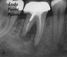
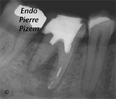
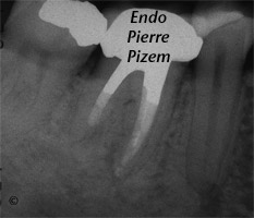
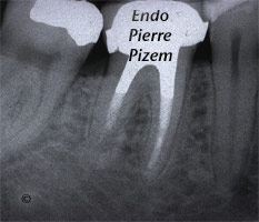
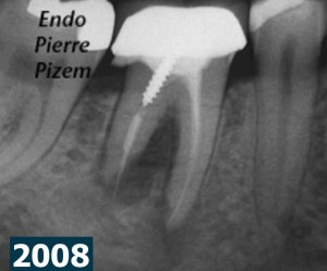
Dental Operative Microscope and Retreatment, Orthograde MTA plugs and root repairs, Root Canal Treatment Pre-Therapy 430646-1 Dental Operative Microscope and Retreatment, Orthograde MTA plugs and root repairs, Root Canal Treatment Per-Therapy 430646-1Dental Operative Microscope and Retreatment, Orthograde MTA plugs and root repairs, Root Canal Treatment Per-Therapy 430646-2  Dental Operative Microscope and Retreatment, Orthograde MTA plugs and root repairs, Root Canal Treatment Per-Therapy 430646-3Dental Operative Microscope and Retreatment, Orthograde MTA plugs and root repairs, Root Canal Treatment Post-Therapy 430646-1Dental Operative Microscope and Retreatment, Orthograde MTA plugs and root repairs, Root Canal Treatment Post-Therapy 430646-2


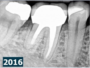
Root canal procedure and periradicular tissu healing 2016-02-21Root canal procedure and periradicular tissue healing 2016-02-21

Case Study Number 430646

External root resorption associated with chronic apical periodontitis altered the shape and position of the foramen through osteoclastic activity, in the x ray images (2008), the modified foramen in distal root is positioned farther from the radiographic apex and gutta percha appears in overextension. A large and circumbscribed radiolucency involves both roots as well as the furcation. This indicates an important periradicular tissues destruction. Tooth mobility level 2 goes along with this tissue loss.

Based both on this 2008 pre operative X ray dental image and clinical findings, should an extraction plus an implant have been an appropriate decision? most certainly  not in my book, time has proven otherwise. Still, up until nowadays, this case is definitely a controversial one, meaning that it is possible for different practitioners to prognosticate endodontic success (very few among practioners) or failure with a great amount of disparity.

Endododontic retreatment and MTA root-end fillings have been performed with a Zeiss Pro Ergo Microscope in september 2008. Last displayed X ray film on this post shows an 8 years post operative clinical outcome. Radiographic examination shows a complete regeneration of periradicular tissues and a resorptive defect healing. An implant would not have been indicated in this case.

As stated by John I. Ingles: “The practicing dentist should not be cited for faulty judgment when even the so-called experts tends to disagree on prognosis… All in all, one must ultimately develop confidence in one’s own abilities. Being able to practice using a great variety of techniques and not being “married” to a single approach in every case will greatly enhance one’s capabilities. And on this is based good prognosis, the result of skill, knowledge, and self confidence.”

Read more about MTA Precision placement with the microscope (.pdf)

Filed Under: Dental operating microscope (D.O.M.) assisted R.C.T., Dental operative microscope and retreatment, Extreme endo clinical cases, NEW CASES, Orthograde MTA plugs and root repairs, Post endodontic treatment outcomes, Regeneration of the periradicular tissues, Resorptive defects healings, Dealing with casted and machined posts removal Tagged With: endodontic file, endodontic revision, external root resorption, microendodontics, Mta, mta apical obturation, Opmi Proergo microscope, orthograde mta placement, posterior mta plug, prognosis, regeneration of periradicular tissues, Retreatment, root canal procedure, root end filling material

Reader Interactions

Comments

  1. Daniel Flynn says

    February 21, 2016 at 6:48 pm

    Beautiful healing Pierre. Congratulations on a fantastic result

    Reply
    • Dr Pierre Pizem says

      February 21, 2016 at 6:52 pm

      Thanks Daniel

      Reply

Leave a Reply Cancel reply

Your email address will not be published. Required fields are marked *

Primary Sidebar

Quality Endodontics

 25+ Years of Experience
in Root Canal Treatment

 300+ Clinical Cases
Published for dentists

 Conveniently Located
Near Metro Vendôme

Call for an appointment:

(514) 667-9541

Clinical cases by categories:

Pierre Pizem Endodontics

A little message from Dr Pizem:

« I understand how stressful going to the dentist could be for you, the patient. So let me reassure you. I’ve built my entire reputation on my professionalism and the passion I pour into my craft. I will not compromise on equipment quality, nor on the efforts necessary to restore your teeth. In the end, my greatest satisfaction is to see you fully recover your dental health, so you may grab big bites at life again. »

Ready to start your treatment?

In order to avoid risks of further complications, we recommend you book a visit as early as you can. However, if you're already feeling pain or urgent symptoms, we'll make every attempt to see you today.

Get in touch

Footer

Contact

514-667-9541

REQUEST APPOINTMENT

Monday
8:30 am - 4:30 pm
Tuesday
8:30 am - 4:30 pm
Wednesday
8:30 am - 4:30 pm
Thursday
8:30 am - 4:30 pm
Friday
8:30 am - 4:30 pm
Sat. Sun.
Closed

Clinic info

  • Home
  • About
  • Dr Pizem
  • Publications
  • Referring Dentists
    • Refer a patient
    • Clinical cases
  • Français

Patient info

  • Root Canal Therapy
  • General Dentistry
  • Endodontic Retreatment
  • FAQ New Patients
  • Tooth Saving Tips
5025 Sherbrooke St. W, Suite 330A
Montreal (QC) H4A 1S9
Near Metro Vendôme       Directions

Copyright © 2021 Endomontreal

Return to top