


Case Study Number 430646
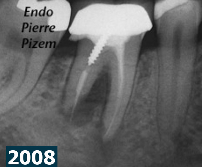
External root resorption associated with chronic apical periodontitis altered the shape and position of the foramen through osteoclastic activity, in the x ray images (2008), the modified foramen in distal root is positioned farther from the radiographic apex and gutta percha appears in overextension. A large and circumbscribed radiolucency involves both roots as well as the furcation. This indicates an important periradicular tissues destruction. Tooth mobility level 2 goes along with this tissue loss.
Based both on this 2008 pre operative X ray dental image and clinical findings, should an extraction plus an implant have been an appropriate decision? most certainly not in my book, time has proven otherwise. Still, up until nowadays, this case is definitely a controversial one, meaning that it is possible for different practitioners to prognosticate endodontic success (very few among practioners) or failure with a great amount of disparity.
Endododontic retreatment and MTA root-end fillings have been performed with a Zeiss Pro Ergo Microscope in september 2008. Last displayed X ray film on this post shows an 8 years post operative clinical outcome. Radiographic examination shows a complete regeneration of periradicular tissues and a resorptive defect healing. An implant would not have been indicated in this case.
As stated by John I. Ingles: “The practicing dentist should not be cited for faulty judgment when even the so-called experts tends to disagree on prognosis… All in all, one must ultimately develop confidence in one’s own abilities. Being able to practice using a great variety of techniques and not being “married” to a single approach in every case will greatly enhance one’s capabilities. And on this is based good prognosis, the result of skill, knowledge, and self confidence.”
Read more about MTA Precision placement with the microscope (.pdf)
Beautiful healing Pierre. Congratulations on a fantastic result
Thanks Daniel