• Skip to primary navigation
  • Skip to main content
  • Skip to primary sidebar
  • Skip to footer

  (514) 667-9541

  REQUEST APPOINTMENT

HOME

ABOUT

DR PIZEM

FRANÇAIS

Endomontreal Root Canal Clinic logo

ENDOMONTREAL

Dr Pizem Advanced Endodontics Clinic

  • Advanced Root Canal Treatment
    • Symptoms & Procedure
    • Why Micro Endodontics?
  • General Dentistry
  • Patients info
    • Frequently asked questions (FAQ)
    • Having a failed root canal treatment?
    • Treatment sequence at our clinic
    • Tooth saving tips
  • For Dentists
    • Refer a patient
    • Clinical cases
Contact Us

Microendodontics with Zeiss OPMI PRO Ergo Operative Microscope: Striving to Find a Pathway to the Apices and to Clean a “C” Shape RC system.

October 28, 2011 By Dr Pierre Pizem Leave a Comment

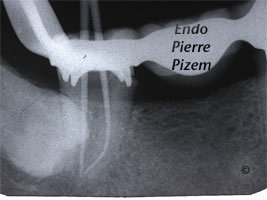
The name comes from the letter “C” shape appearance of a very large isthmus in the pulp chamber floor when viewed from above. This isthmus or groove is the result of the merging of some or all of the root canals at the cervical area near the pulp chamber floor. Incidence is 2,7% in Caucasian and up to 13% in asian population. Pre operative X Ray dental film shows a blurred image of the canal system, canals are not visible and pulp chamber is almost not visible. Looking at these features it may not be possible to diagnose a C shape canal but we must suspect either this canal configuration or severe fibrosis/calcification.

“C” shape canals are a real challenge to preparation and may cause technical complications such as transportation, steps, stripping with perforation in the thin wall area or blockage of the canal.

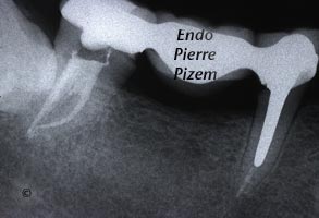
This procedure requires a full understanding of this anatomy to prepare an optimal access cavity to pulp chamber through a PFM abutment,  to know where to look for the root canal entries and to be cautious about the thin wall area. This endodontic procedure also requires much more operating chair time for debridment. No rapid techniques does exist to shape clean and fill those peculiar root canal shapes. This specific endodontic procedure also justifies the use of a dental operative microscope to better see what we are doing.

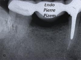
Atypical Canal Configurations, 'C' Shaped Canal System, Root Canal Treatment Pre-Therapy 319947-1  Atypical Canal Configurations, 'C' Shaped Canal System, Root Canal Treatment Per-Therapy 319947-1  Atypical Canal Configurations, 'C' Shaped Canal System, Root Canal Treatment Post-Therapy 319947-1

Endodontist. Case Study Number 319947.

This 3D video of a “C” shaped second mandibular molar from the rootcanalanatomyprojectblogspot.com displays the complex anatomy of such a root canal system. In just a few second the video gives a better understanding on how difficult the cleaning and filling tasks of a “C” shape root canal may be.

Last september, a new generation of endodontic file has been presented at the CAE meeting in Quebec city-Canada by Dr Zvi Metzger, Professor and Chair Department of Endodontology school of Dental Medicine at Tel Aviv University. Although at the moment, the Self Adjusting File System (SAF System) is not yet readily available everywhere in Canada, this innovative endodontic file adapts to root canal shape thus, may represent in a near future, a valuable approach to more efficient debridement in C shape canals and a safer way to address thin wall section that is always present in this anatomical variation. Here is a promotional video showing how it works.

Filed Under: Atypical canal configurations, Extreme endo clinical cases, General, NEW CASES, 'C' shaped canal system Tagged With: 3d video, c shape root canal, cae, cleaning, Dental operative microscope, endodontic file, endodontic files, Endodontics, Endodontist, filling, Gutta percha, metzger zvi, microendodontics, Microscope, opmi proergo, Opmi Proergo microscope, pierre, pizem, procedure, pulp canal sealer, Root canal treatment, saf, saf system, shaping, system

Reader Interactions

Leave a Reply Cancel reply

Your email address will not be published. Required fields are marked *

Primary Sidebar

Quality Endodontics

 25+ Years of Experience
in Root Canal Treatment

 300+ Clinical Cases
Published for dentists

 Conveniently Located
Near Metro Vendôme

Call for an appointment:

(514) 667-9541

Clinical cases by categories:

Pierre Pizem Endodontics

A little message from Dr Pizem:

« I understand how stressful going to the dentist could be for you, the patient. So let me reassure you. I’ve built my entire reputation on my professionalism and the passion I pour into my craft. I will not compromise on equipment quality, nor on the efforts necessary to restore your teeth. In the end, my greatest satisfaction is to see you fully recover your dental health, so you may grab big bites at life again. »

Ready to start your treatment?

In order to avoid risks of further complications, we recommend you book a visit as early as you can. However, if you're already feeling pain or urgent symptoms, we'll make every attempt to see you today.

Get in touch

Footer

Contact

(514) 667-9541

REQUEST APPOINTMENT

Monday
8:30 am - 4:30 pm
Tuesday
8:30 am - 4:30 pm
Wednesday
8:30 am - 4:30 pm
Thursday
8:30 am - 4:30 pm
Friday
8:30 am - 4:30 pm
Sat. Sun.
Closed

Clinic info

  • Home
  • About
  • Dr Pizem
  • Publications
  • Referring Dentists
    • Refer a patient
    • Clinical cases
  • Français

Patient info

  • Root Canal Therapy
  • General Dentistry
  • Endodontic Retreatment
  • FAQ New Patients
  • Tooth Saving Tips
5025 Sherbrooke St. W, Suite 330A
Montreal (QC) H4A 1S9
Near Metro Vendôme       Directions

Copyright © 2021 Endomontreal

Return to top