• Skip to primary navigation
  • Skip to main content
  • Skip to primary sidebar
  • Skip to footer

  514-667-9541

  REQUEST APPOINTMENT

HOME

ABOUT

DR PIZEM

FRANÇAIS

ENDOMONTREAL

ENDOMONTREAL

Dr Pizem Advanced Endodontics Clinic

  • Advanced Root Canal Treatment
    • Symptoms & Procedure
    • Why Micro Endodontics?
  • General Dentistry
  • Patients info
    • Frequently asked questions (FAQ)
    • Having a failed root canal treatment?
    • Treatment sequence at our clinic
    • Tooth saving tips
  • For Dentists
    • Refer a patient
    • Clinical cases
Contact Us

Carl Zeiss OPMI PROergo Insured Enough Visual Accuracy to Prevent a Missed Apical Split in a Calcified Mesial Root.

March 29, 2012 By Dr Pierre Pizem Leave a Comment

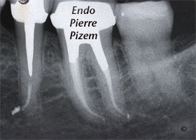
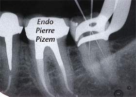
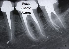
Atypical Canal Configurations, Type V, Root Canal Treatment Pre-Therapy 197337-1  Atypical Canal Configurations, Type V, Root Canal Treatment Per-Therapy 197337-1  Atypical Canal Configurations, Type V, Root Canal Treatment Post-Therapy 197337-1


Endodontist. Case Study Number 197337

The recent addition of dental operative microscope (DOM) to endodontic therapy can allow better visualization and management of the intricate morphology of the root canal system during endodontic procedures through magnification and greatly improved high intensity lighting. Dental Microscope typically magnifies in the 4X to 25X range. The other commonly used magnification aide, through lens eyeglass mounted surgical telescopes, provides 2.5X to 4.5X magnification. 

We have been presented with this second mandibular molar that has only two canal entries on pulpal chamber floor. At first sight one could have easily concluded the presence of only two canals. In fact, the mesial root has a Vertucci’s type 5 canal configuration. A Vertucci type V pulp space configuration can be described as follow: One canal leaves the pulp chamber and divides short of the apex into two separate distinct canals with two distinct foramina (1-2). Without magnification the root canal apical “split” could have been under seen, treating one branch out of two and leaving  pulp tissue inside the other branch.

Surgical operating microscopes have a steep learning curve and require training, as well as patience and practice to master. Still this piece of equipment and the learning effort it implies is well worth it since cases that once seemed impossible can now be treated with a high degree of confidence and clinical success. 

As the saying goes:”A picture is worth a thousand words”, Click here to have a look at what can be seen at an operative field under magnified observation (10X to 25X range). 

Filed Under: Atypical canal configurations, Curved canals, Extreme endo clinical cases, General, NEW CASES, Type V, What's new?, D.O.M. versus partially calcified systems Tagged With: Dental operative microscope, endodontic microscope, endodontic procedure, Endodontist, microendodontics, Microscope, Opmi Proergo microscope, Protaper dentsply, root canal apical split, root canal procedure, Root canal system calcifications, Root canal treatment, surgical operative microscope, vertucci type V root canal configuration

Reader Interactions

Leave a Reply Cancel reply

Your email address will not be published. Required fields are marked *

Primary Sidebar

Quality Endodontics

 25+ Years of Experience
in Root Canal Treatment

 300+ Clinical Cases
Published for dentists

 Conveniently Located
Near Metro Vendôme

Call for an appointment:

(514) 667-9541

Clinical cases by categories:

Pierre Pizem Endodontics

A little message from Dr Pizem:

« I understand how stressful going to the dentist could be for you, the patient. So let me reassure you. I’ve built my entire reputation on my professionalism and the passion I pour into my craft. I will not compromise on equipment quality, nor on the efforts necessary to restore your teeth. In the end, my greatest satisfaction is to see you fully recover your dental health, so you may grab big bites at life again. »

Ready to start your treatment?

In order to avoid risks of further complications, we recommend you book a visit as early as you can. However, if you're already feeling pain or urgent symptoms, we'll make every attempt to see you today.

Get in touch

Footer

Contact

514-667-9541

REQUEST APPOINTMENT

Monday
8:30 am - 4:30 pm
Tuesday
8:30 am - 4:30 pm
Wednesday
8:30 am - 4:30 pm
Thursday
8:30 am - 4:30 pm
Friday
8:30 am - 4:30 pm
Sat. Sun.
Closed

Clinic info

  • Home
  • About
  • Dr Pizem
  • Publications
  • Referring Dentists
    • Refer a patient
    • Clinical cases
  • Français

Patient info

  • Root Canal Therapy
  • General Dentistry
  • Endodontic Retreatment
  • FAQ New Patients
  • Tooth Saving Tips
5025 Sherbrooke St. W, Suite 330A
Montreal (QC) H4A 1S9
Near Metro Vendôme       Directions

Copyright © 2021 Endomontreal

Return to top