• Skip to primary navigation
  • Skip to main content
  • Skip to primary sidebar
  • Skip to footer

  514-667-9541

  REQUEST APPOINTMENT

HOME

ABOUT

DR PIZEM

FRANÇAIS

ENDOMONTREAL

ENDOMONTREAL

Dr Pizem Advanced Endodontics Clinic

  • Advanced Root Canal Treatment
    • Symptoms & Procedure
    • Why Micro Endodontics?
  • General Dentistry
  • Patients info
    • Frequently asked questions (FAQ)
    • Having a failed root canal treatment?
    • Treatment sequence at our clinic
    • Tooth saving tips
  • For Dentists
    • Refer a patient
    • Clinical cases
Contact Us

How the Use of Opmi PROergo Dental Operative Microscope Prevented an Unnecessary Molar Extraction. An Endodontist Case Study.

November 7, 2012 By Dr Pierre Pizem Leave a Comment

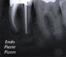
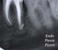
Atypical Canal Configurations, Type V, Root Canal Treatment Pre-Therapy 524236-1    Atypical Canal Configurations, Type V, Root Canal Treatment Per-Therapy 524236-1    Atypical Canal Configurations, Type V, Root Canal Treatment Post-Therapy 524236-1

An intricate root canal procedure. Case study number 524236

Two successive root canal procedure attemps failed on this mandibular molar. In the past three years patient had to take many courses of antibiotics to control pain and swelling. Patient is unable to chew on that tooth. An extraction and an implant supported crown has been suggested but patient wants to keep his own tooth despite all this.

Clinical examination shows vestibular swelling, probing does not show any narrow deep pocket. On radiographic examination filling material is overextended in distal root and a large bone rarefaction area is present on distal root tip.

Retreatment of the root canal on distal root is suggested because an untreated  fourth canal is suspected. Once  filling material has been removed no extra canal could be found, instead, with the help of high magnification an apical delta or Vertucci’s type V pulp space configuration could be noticed. A Vertucci type V pulp space configuration can be described as follow: One canal leaves the pulp chamber and divides short of the apex into two separate distinct canals with distinct foramina (1-2). According to Vertucci’s study in 1984 on a 100 mandibular molars sample, the type V configuration in distal root, occurred only in 8% of the teeth examined.

Consequently the untreated branch is filled with necrotic pulp and bacteria releasing toxins into the surrounding bone area. An ISO files number 08 is inserted into the untreated branch, then, a NiTi file is also inserted into the retreated branch through the same canal entry and a second Xray is taken. The second Xray clearly displays the apical split in last apical third of distal single canal. Each part of the split in distal root has been individually cleaned and shaped. NiTi files allowed us to follow both curved branches. Root canals have been filled with calcium Hydroxide and patient came back 8 days later to have those filled with Pulp Canal Sealer and gutta percha. (Lateral and vertical condensation). Last (Angulated) X ray to the right shows the two branches after final obturation.

Tooth symptoms have subsided shortly after calcium hydroxide have been inserted into the root canal system. Two months have passed and tooth is still symptoms free. Being able to get magnification and bring illumination to the root canal tip allowed for that tooth to be preserved. Patient was told to protect his tooth with cusp coverage.

Filed Under: Atypical canal configurations, Dental operative microscope and retreatment, Extreme endo clinical cases, Type V, What's new? Tagged With: apical delta, endodontic procedure, necrotic pulp, pulp canal sealer, Root canal, root canal apical split, root canal procedure, root canal split, vertucci type V root canal configuration

Reader Interactions

Leave a Reply Cancel reply

Your email address will not be published. Required fields are marked *

Primary Sidebar

Quality Endodontics

 25+ Years of Experience
in Root Canal Treatment

 300+ Clinical Cases
Published for dentists

 Conveniently Located
Near Metro Vendôme

Call for an appointment:

(514) 667-9541

Clinical cases by categories:

Pierre Pizem Endodontics

A little message from Dr Pizem:

« I understand how stressful going to the dentist could be for you, the patient. So let me reassure you. I’ve built my entire reputation on my professionalism and the passion I pour into my craft. I will not compromise on equipment quality, nor on the efforts necessary to restore your teeth. In the end, my greatest satisfaction is to see you fully recover your dental health, so you may grab big bites at life again. »

Ready to start your treatment?

In order to avoid risks of further complications, we recommend you book a visit as early as you can. However, if you're already feeling pain or urgent symptoms, we'll make every attempt to see you today.

Get in touch

Footer

Contact

514-667-9541

REQUEST APPOINTMENT

Monday
8:30 am - 4:30 pm
Tuesday
8:30 am - 4:30 pm
Wednesday
8:30 am - 4:30 pm
Thursday
8:30 am - 4:30 pm
Friday
8:30 am - 4:30 pm
Sat. Sun.
Closed

Clinic info

  • Home
  • About
  • Dr Pizem
  • Publications
  • Referring Dentists
    • Refer a patient
    • Clinical cases
  • Français

Patient info

  • Root Canal Therapy
  • General Dentistry
  • Endodontic Retreatment
  • FAQ New Patients
  • Tooth Saving Tips
5025 Sherbrooke St. W, Suite 330A
Montreal (QC) H4A 1S9
Near Metro Vendôme       Directions

Copyright © 2021 Endomontreal

Return to top