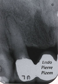
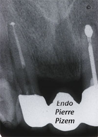
Dental operating microscope assisted root canal procedure on a completely stenosed canal system (calcified canal).
Endodontist. Case Study Number 506712
In order to be preserved and used as an abutment for a fixed bridge replacement, this tooth must have a casted post space preparation in its coronal third. Thus apical endodontic surgery cannot be considered as the best option for the patient.
Pre operative condition:
- Canal is not visible on dental X ray film until last few millimeters because the root canal anatomy system does not begin before last few millimeters, this means an extremely narrow canal diameter for the practioner to locate in last apical third of root. Remaining canal diameter can be 3 times smaller than a single strand of human hair diameter. Remaining within tooth long axis when accessing canal entry is of the utmost importance not to create a iatrogenic perforation.
- Two previous failed attempts to locate tooth single canal entry, this means complete loss of landmarks when looking through dental operative microscope lens to find it
- Number 12 tooth is a 12X21 bridge abutment, this means loss of external landmarks to locate canal entry
- Dentine shade composite completely fills up the access cavity, this means even more challenge, when drilling to expose canal entry, not to create additional tooth substance loss (thus increasing tooth weakness.
Tooth survival relies solely on endodontic procedure success, if canal cannot be found thus treated, tooth cannot be preserved.
While being a less straightforward approach than implant therapy, this successful endodontic procedure outcome strongly suggests that, nowadays, a complex root canal retreatment does not necessarily have to mean extraction and replacement by a dental implant.
Surgical operating microscopes have a steep learning curve and require training, as well as patience and practice to master. Still this piece of equipment and the learning effort it implies is well worth it since cases that once seemed impossible can now be treated with a high degree of confidence and clinical success.


Dear Dr. Pizem,
I’am writing you from Belgrade, Serbia. I hope you will find some time to answer me since I find you one of the world leaders in endodontics.
The problem I’am dealing with is calcified cuspid. I had difficulty locating the canal but I menage to do it, however the canal was not patent to the apex. I used precurved k file no 10 with watch winding motion and lot of edta but instrument still binds at the begining of the apical third.
The problem is that on xray I cannot identify canal apical from this binding point. My question is: is there any way to establish canal patency through entire length in cases like this (when canal is obviously calcified on x ray in the apical third, mostly seen in older patients) or is the only way cleaning, shapping and obturating to that point?
Since I am often dealing with calcified canalas and I saw a lot of interesting cases about calcifed root canals at your web site I would be very greatfull to hear about your experience regarding this topic.
Best Regards from Serbia
Ivan Mirovic DDS
Dear Dr Mirovic,
You have already achieved a wonderful work with your no 10 file used in a watch winding motion. You have succeeded with lateral incisor and there is so little to achieve same result in this calcified cusp. Bravo!
In order to complete full debridement and shaping I would only suggest the use of a 06 or 08 precurved C file from Maillefer or other good brand mark in conjunction with a chelating agent. Scout in order to get this famous “catch”, once you’ve got it HOLD IT! and reduce amplitude of your motion keeping the tip of your instrument in that same orientation as much as you can. It will lead you to the foramina. The rest of shaping is routine.
Do not worry too much about not seeing canal on X ray, it is there all right. Rely on on your tactile sense now, you have to feel the “catch”
All the best from Canada
Dear Dr Pizem,
I manage to do it! I followed your instructions and used no8 k file highly curved at the tip ( 90 degrees) and passed easily. Thanks a lot for great help!
All the best