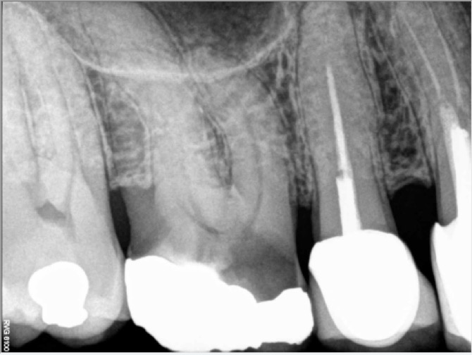

The patient urged my referring dentist to save his own natural tooth even though it was badly decayed and it presented with an uncomplicated crown fracture on its lingual side. Despite its apparent questionable restorability on pre operative Xray, the tooth is very long and its periodontal status is good so we accepted to perform both the root canal procedure and the post and core build up.
RCT procedure on this maxillary molar has been a time consuming one mostly due to its calcified root canal system and its 26mm length. MB2 root canal entry has been located with the help a dental operating microscope high magnification along with a coaxial Xenon illumination.
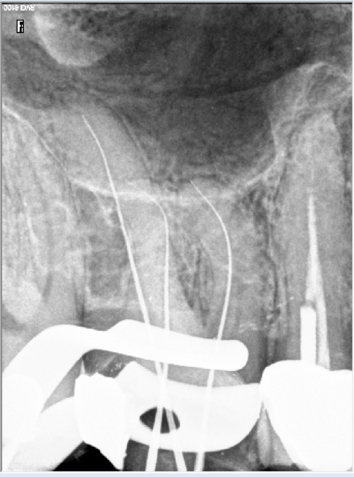
Armamentarium: Zeiss’s OPMI PROergo Dental Operative Microscope , 556 straight cross cut carbide tungsten bur, Ultrasonic tips (BUC 3), RC Prep, many Mani K files (envelop of motion), ProTaper Universal, 6% NaOCl, PUI, Pulp Canal Sealer + W.V.C.
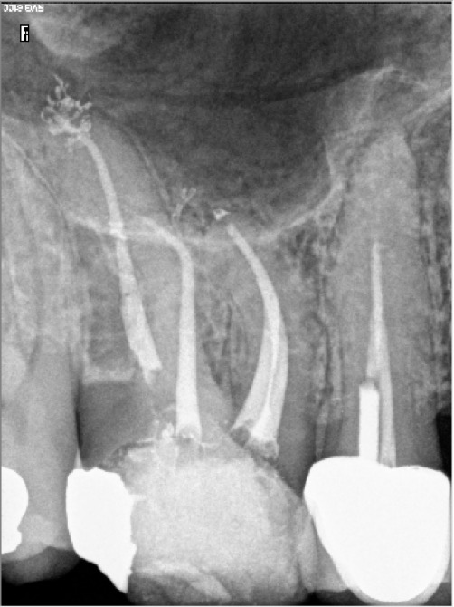
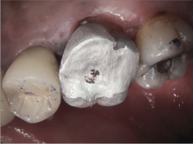
Tooth has been restored with a FlexiPost (cemented with RelyX luting cement) and an amalgam core build up. (Slight excess of amalgam seen on post operative X ray on palatal aspect of the tooth has been removed prior to sending patient back to his own dentist for crowning)
Leave a Reply