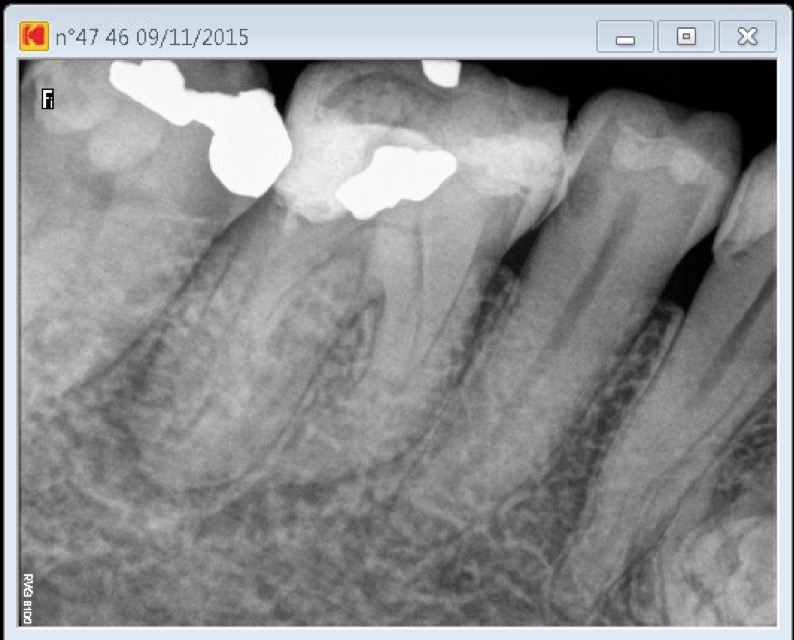
Patient presented to our office with an AAA.
To complete the root canal procedure on this dilacerated tooth, access opening has been performed with a combination of a 556 cross cut and a round tungsten carbide bur (Friction Grip). Operative field observation has been enhanced with high magnification and coaxial xenon lamp illumination (Carl Zeiss OPMI PROergo dental operative microscope). Dystrophic calcifications have been removed from pulp chamber with ultrasonic diamond coated tips in order to expose both the distal root canal entries location. An interim calcium hydroxide dressing has been inserted and patient had to come back in order to gain control over the acute inflammation prior to filling the root canals. (Patient has been advised concerning cavities on the adjacent teeth).
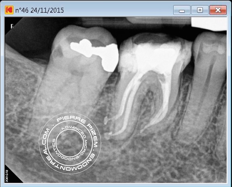
Dilacerations instrumentation have been performed with Mani stainless K files and ProTaper Universal (Dentsply). Chelator: RC Prep. PUI Irrigation: NaOCl 5%. Ultrasonic tip: BUC One from Spartan. P5 (Dentsply) Sealer used: Pulp Canal Sealer. Obturation technique WVC.
X Ray Sensors used: CareStream Kodak 6100.
Great case as usual!