Anatomical variations must be considered at the beginning of the root canal treatment. Even though at this point, accurate preoperative radiographs are considered essential giving indications to clinicians as to the number of roots and canals that exist in a tooth, adequate examination of pulp chamber floor with the help of magnification and co axial illumination may give to clinician much more extensive information about number, position of pulp canal orifices and wall anatomy.
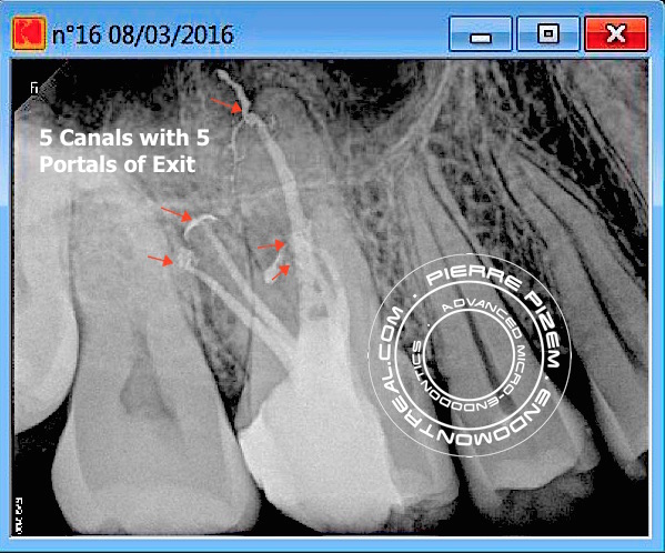
In this rare anatomical variation 3 canal entries could be located: 1 in palatal root, 2 in distal root, and only 1 in the mesio vestibular root. The mesiovestibular single root canal entry did split into two canals at a depth of 2mm below the pulp chamber floor level.
Leave a Reply