• Skip to primary navigation
  • Skip to main content
  • Skip to primary sidebar
  • Skip to footer

  (514) 667-9541

  REQUEST APPOINTMENT

HOME

ABOUT

DR PIZEM

FRANÇAIS

Endomontreal Root Canal Clinic logo

ENDOMONTREAL

Dr Pizem Advanced Endodontics Clinic

  • Advanced Root Canal Treatment
    • Symptoms & Procedure
    • Why Micro Endodontics?
  • General Dentistry
  • Patients info
    • Frequently asked questions (FAQ)
    • Need a root canal revision?
    • Treatment sequence at our clinic
    • Tooth saving tips
  • For Dentists
    • Refer a patient
    • Clinical cases
    • Implants vs Endodontics
Contact Us

A Huge Radiographic “J” Shape Lesion, Does the Tooth Has a Vertical Root Fracture? a Periodontal Infection? an Endodontic Infection?

You are here: Home / Dental operating microscope (D.O.M.) assisted R.C.T. / A Huge Radiographic “J” Shape Lesion, Does the Tooth Has a Vertical Root Fracture? a Periodontal Infection? an Endodontic Infection?

March 31, 2011 By Dr Pierre Pizem Leave a Comment

Dental operating microscope (D.O.M.) assisted R.C.T., Dental operative microscope and retreatment, Root Canal Treatment Pre Therapy 456147-1

Case Study Number 456147

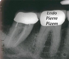
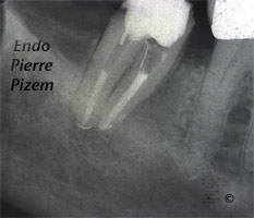
We have been presented with this mandibular second molar that has a periapical lucency which also has a periodontal component. Patient is experiencing pain. This radiographic image also shows a huge radiographic J-shaped lesion that may be indicative of a vertical root fracture in the mesial aspect of the mesial root.

Are we dealing with a periodontal infection? And, if this is the case, such a periradicular tissues loss would command an extraction of the tooth.

Are we dealing with a fracture? And, if this is the case, tooth extraction would also be the treatment of choice.

Are we dealing with an endodontic infection? If this is the case an endodontic revision (endodontic retreatment) would suffice to preserve this tooth.

The decision of extracting or saving that tooth should be based on a foundation of sound diagnosis.

One of the primary fact that needs to be established in distinguishing endo/perio lesion is the pulp status. Since there is not much pulp left in that case, pulp testing won’t help much.

Periodontal probing is the next important measurement in determining if wether this tooth has an endodontic or periodontic infection. Probing shows no wide or narrow pocket on buccal neither on lingual (and on distal) aspects of this tooth, at this stage, periodontal infection might be ruled out.

The remaining question is: “Are we dealing with a fracture or an endodontic infection?”
A deep narrow periodontal pocket would indicate a vertical root fracture and the quiz would end right there. The tooth would have to be extracted.The problem is that this defect, if it does exist, might be located just beneath a wide and tight mesial contact point and probing with accuracy this area is not that obvious. Hence the final question cannot be answered without any doubt by probing alone.

One option would be to remove the crown to allow for a direct access to the potential defect. But then, if no deep narrow periodontal pocket exist, the clamp for the rubber dam as well as tooth protection from vertical forces would require a provisional crown to be installed. Even so, probing a deep narrow periodontal pocket is not an absolute waranty that this defect is in deed a crack. It might also be a desmodontal sinus tract. Hence,drilling through the existing crown to access mesials root canals and a direct observation under magnifying lenses of a microscope seemed to be the appropriate way of confirming the presence or absence of a vertical root fracture. (But, then again if crack would have been located in the apical third of root canal system, microscope lenses would not have been of much help either…)

Dental operating microscope (D.O.M.) assisted R.C.T., Dental operative microscope and retreatment, Root Canal Treatment Per Therapy 456147-1
Dental operating microscope (D.O.M.) assisted R.C.T., Dental operative microscope and retreatment, Root Canal Treatment Per Therapy 456147-2
Dental operating microscope (D.O.M.) assisted R.C.T., Dental operative microscope and retreatment, Root Canal Treatment Per Therapy 456147-3
Dental operating microscope (D.O.M.) assisted R.C.T., Dental operative microscope and retreatment, Root Canal Treatment Per Therapy 456147-4

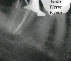
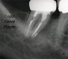
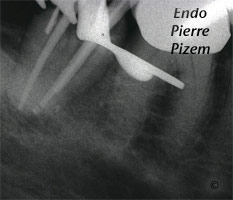
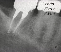
No crack could be observed from within the mesial root canals, the treatment has been completed in two appointments (third, fourth and fifth images) . Following the first appointment which implied material retrieval, irrigation and calcium hydroxyde insertion (Second X ray image), both pain and swelling disappeared. Following Xray image shows a complete periodontal tissue regeneration after one year. Tooth is asymptomatic.

Dental operating microscope (D.O.M.) assisted R.C.T., Dental operative microscope and retreatment, Root Canal Treatment Post Therapy 456147-1

Article on perio endo lesions

Category iconDental operating microscope (D.O.M.) assisted R.C.T.,  Dental operative microscope and retreatment,  Dental operative microscope versus cracks,  Extreme endo clinical cases,  NEW CASES,  Popular Cases,  Post endodontic treatment outcomes,  Regeneration of the periradicular tissues,  The misleading ''J'' Shape lesion

Reader Interactions

Leave a Reply Cancel reply

Your email address will not be published. Required fields are marked *

Primary Sidebar

Quality Endodontics

 30+ Years of Experience
in Root Canal Treatment

 300+ Clinical Cases
Published for dentists

 Conveniently Located
Near Metro Vendôme

Call for an appointment:

(514) 667-9541

Cases with high level of difficulty:

Pierre Pizem Endodontics

A little message from Dr Pizem:

« I understand how stressful going to the dentist could be for you, the patient. So let me reassure you. I’ve built my entire reputation on my professionalism and the passion I pour into my craft. I will not compromise on equipment quality, nor on the effort necessary to restore your teeth. In the end, my greatest satisfaction is to see you fully recover your dental health, so you may grab big bites at life again. »

Ready to start your treatment?

In order to avoid risks of further complications, we recommend you book a visit as early as you can. However, if you're already feeling pain or urgent symptoms, we'll make every attempt to see you today.

Get in touch

Footer

Contact

(514) 667-9541

REQUEST APPOINTMENT

Monday
8:30 am - 4:30 pm
Tuesday
8:30 am - 4:30 pm
Wednesday
8:30 am - 4:30 pm
Thursday
8:30 am - 4:30 pm
Friday
Closed
Sat. Sun.
Closed

Clinic info

  • Home
  • About
  • Dr Pizem
  • Publications
  • Referring Dentists
    • Refer a patient
    • Clinical cases
  • Français

Patient info

  • Root Canal Therapy
  • General Dentistry
  • Endodontic Retreatment
  • FAQ New Patients
  • Tooth Saving Tips
5025 Sherbrooke St. W, Suite 330A
Montreal (QC) H4A 1S9
Near Metro Vendôme       Directions

Copyright © 2025 Endomontreal

Return to top