According to the Canadian Academy of Endodontics, when having to treat a tooth presenting with an exceptionally complicated preoperative condition,
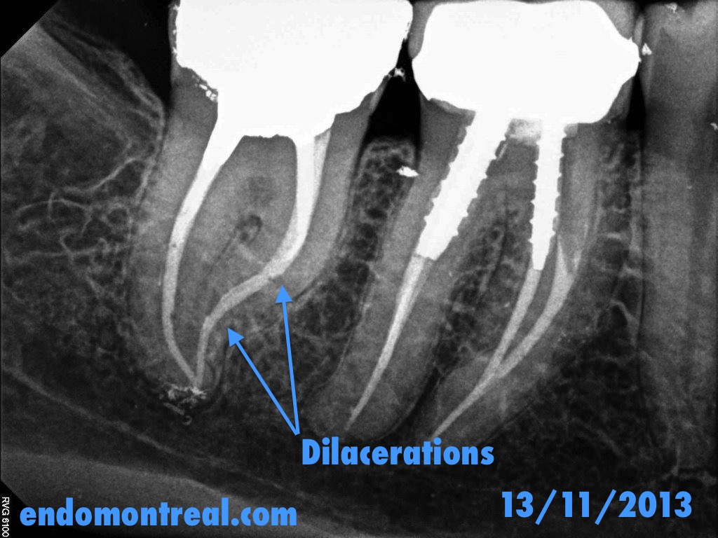
Completely calcified tooth does not necessarily condemn it to extraction
One must not decide a tooth extraction on the sole basis of an x-ray observation. In cases of complete calcification of root canal systems, the
Root Canal Treatment on a Tooth Presenting With a Rare Anatomical Variation
Mandibular first premolar, necrotic pulp, AAA, patient has a gag reflex making it difficult to take a pre op. X ray dental film revealing a peculiar
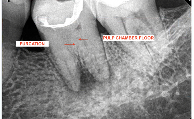
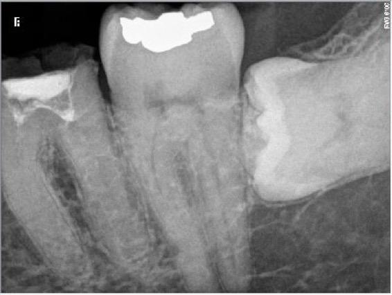
Intricate S Shape Root Canal Procedure on a Calcified Second Mandibular Molar
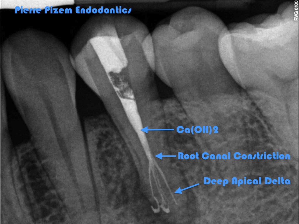
Thinking Out of the Box 20 years Ago to Preserve a Tooth! An Early Ceramic Material Apical Filling
Wide apical foramen, thin canal walls, necrotic pulp would, in year 2000, condemn this tooth and have it replaced by an implant supported
A Painless Root Canal Procedure on a Calcified Tooth. A Difficult and Time Consuming One to Perform
Microendodontics is a Microscope Endodontic Procedure to Preserve an Otherwise Untreatable Tooth
ROOT CANAL PROCEDURE CASE STUDY NUMBER: 542917 Tooth number 16 has to be removed, tooth number 17 becomes a key tooth as an abutment for a fixed
Huge Pulp Stone Removal Under High Magnification in Order to Preserve a Maxillary Molar
&
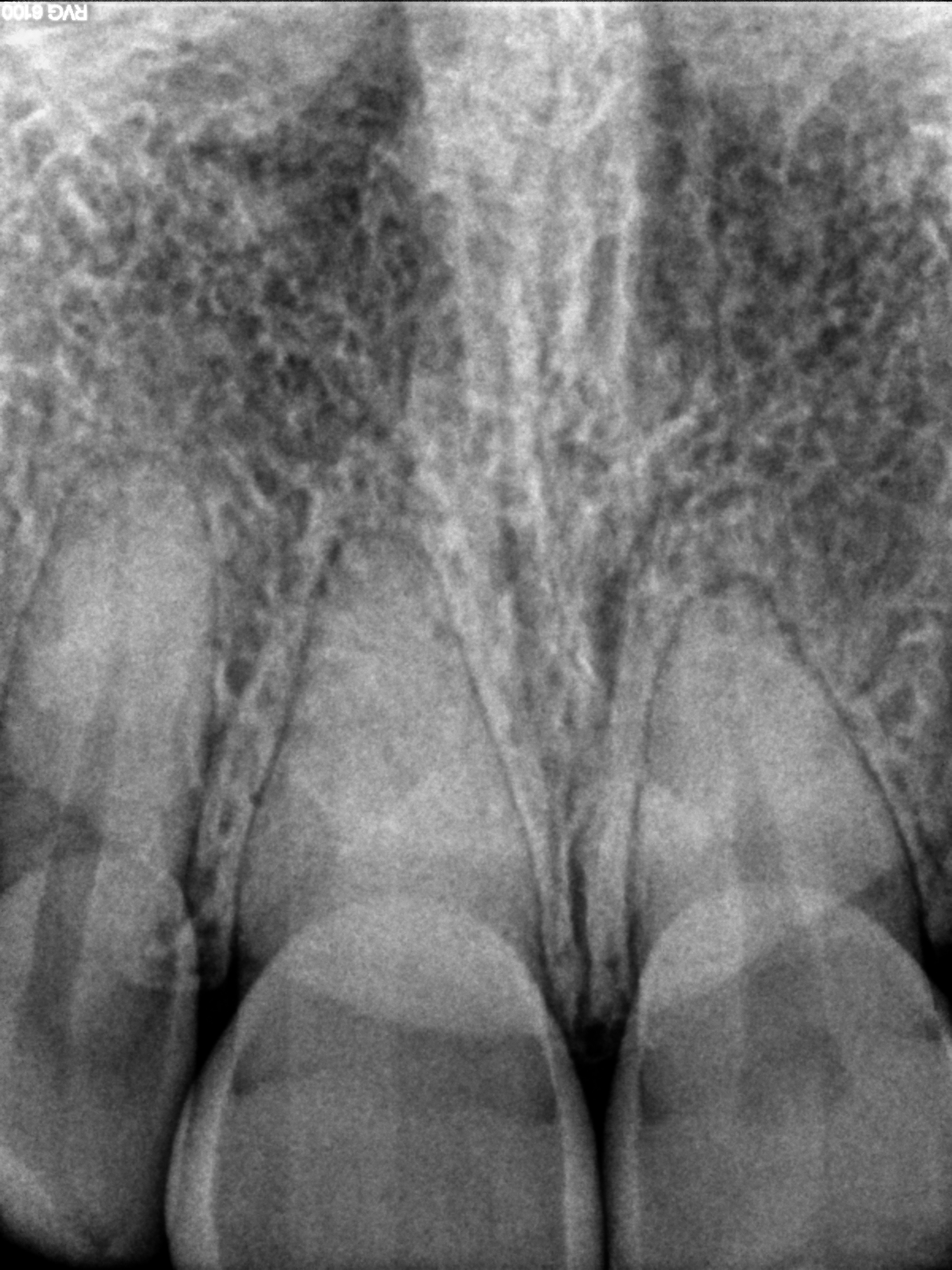
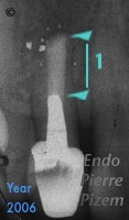
Obliterated Pulp Space VS Dental Operative Microscope
First X Ray image : Pulp space is not visible on tooth number 11 Second X Ray image : Displays a number 06 ISO stainless Steel file from Mani at