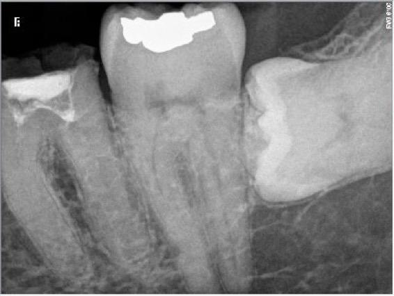
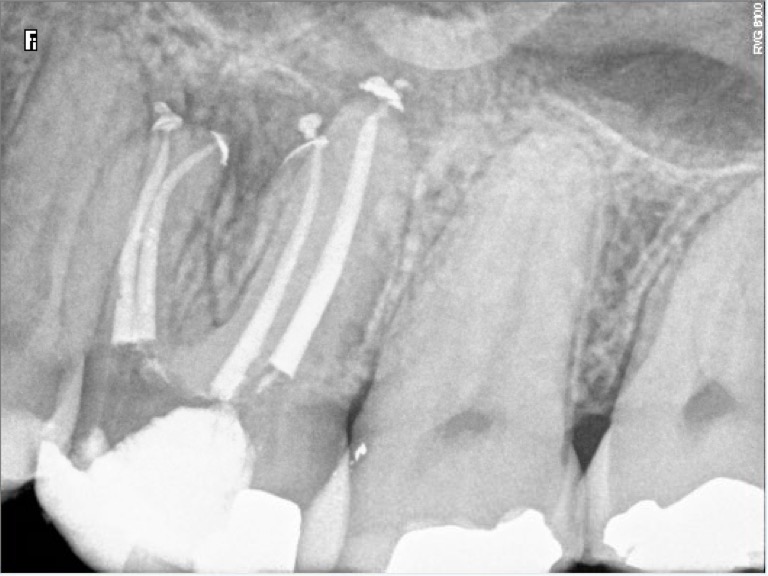
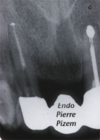
One must not decide a tooth extraction on the sole basis of an x-ray observation. In cases of complete calcification of root canal systems, the
Going the extra mile to secure root canal treatment success
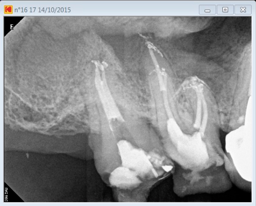
Patient has been referred for root canal treatment on both first and second maxillary molars. Tooth number 16 had necrotic pulp and tooth 17
Too much calcification for a root canal treatment?
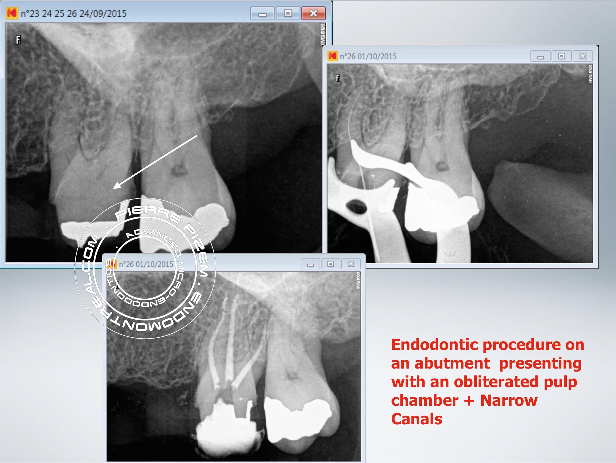
This tooth is a 23XX26 leaking fixed bridge abutment. Prior to bridge replacement it needs a root canal procedure. Performing a root canal
The dental operative microscope does play a key role in difficult root canal treatments
The patient was experiencing a daily constant nagging pain for the last 2 months. Pain increased along with some gum swelling the during last
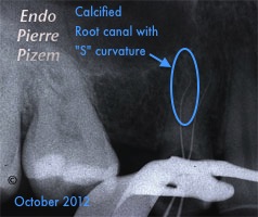
Root Canal Therapy on a Calcified Root Canal With an S Form Curvature (Case 525214)
Endodontic. Case Study Number 525214 Locating both root canal entries without lateraly perforating the root and without destroying to much sound
Removing a Tiny Fractured Instrument Fragment from a Root Canal System.
Mass of embedded pulp stones is still present in pulp chamber and it must be removed to increase both retention and strength of planned core Big
Huge Pulp Stone Removal Under High Magnification in Order to Preserve a Maxillary Molar
&
Opmi Proergo Dental Operative Microscope, a Cutting Edge Technology to Save a Key Tooth. Overcoming an Against All Odds Clinical Pre Operative Condition.
Dental operating microscope assisted root canal procedure on a completely stenosed canal system (calcified canal). Endodontist. Case Study Number
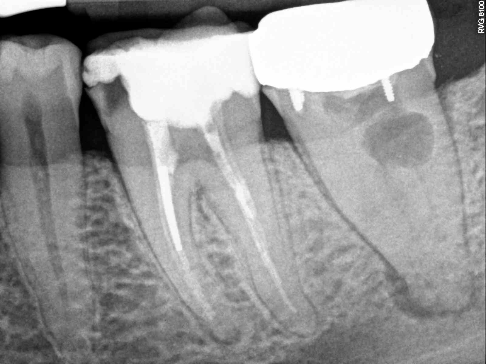
Pitfalls in Endodontic Re-treatment on a Painful Maxillary First Molar
Untreated calcified and contaminated canal segments Hidden (thus untreated) second calcified root canal to locate and treat Sargenti Paste filling

