Endodontist. Root Canal Procedure. Case Study Number 49821617
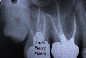
Note on the post operative Xray dental film, the dilacerated apical curves in both vestibular roots on second maxillary molar and disto vestibular root of first maxillary molar. The values of those root canal curvature radius based on three mathematical points are all below 4 mm, these are very small radius. Thus, those cuvatures can be defined as severe. For the endodontist, choosing the right endodontic file is of the utmost importance in order to avoid damaging consequences such as: apical transportations, loss of working length, zip and perforations and fracture of instruments. Calcified canals are making the procedure even more difficult.
An interesting point has been raised by Drs Pruett, Clement and Carnes affiliated with the Department of Endodontics/Dental School of University of Texas Health Science Center at San Antonio:
These results indicate that, for nickel-titanium, engine-driven rotary instruments, the radius of curvature, angle of curvature, and instrument size are more important than operating speed for predicting separation.
Much more can be learned on small curvature radius by reading the following article: Method for determination of root curvature radius using cone beam computed tomography images
Carlos Estrela, Mike Reis Bueno, Manoel Damiao Sousa Neto, Jesus Djalma Pécora Braz Dent J (2008) 19(2): 114-118 ISSN 0103-6440


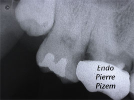
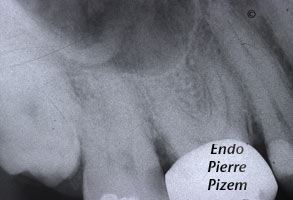
[…] a maxillary second molar presenting a partial mineralization of it’s root canal system. This case report in microendodontics number 49821617 displays an intricate procedure on a mineralised root canal systems with a very small radius […]