• Skip to primary navigation
  • Skip to main content
  • Skip to primary sidebar
  • Skip to footer

  (514) 667-9541

  REQUEST APPOINTMENT

HOME

ABOUT

DR PIZEM

FRANÇAIS

Endomontreal Root Canal Clinic logo

ENDOMONTREAL

Dr Pizem Advanced Endodontics Clinic

  • Advanced Root Canal Treatment
    • Symptoms & Procedure
    • Why Micro Endodontics?
  • General Dentistry
  • Patients info
    • Frequently asked questions (FAQ)
    • Need a root canal revision?
    • Treatment sequence at our clinic
    • Tooth saving tips
  • For Dentists
    • Refer a patient
    • Clinical cases
    • Implants vs Endodontics
Contact Us

Calcified Pulp Chamber with Two Superimposed Floors: A Clinical Approach

You are here: Home / CBCT assisted Root Canal Treatment / Calcified Pulp Chamber with Two Superimposed Floors: A Clinical Approach

February 29, 2020 By Dr Pierre Pizem Leave a Comment

Calcified root canal system provide an endodontic treatment challenge. The introduction of new technologies has increased the predictability of these treatments and, consequently, their success rates. This article describes one case with a clinical treatment strategy using high technology for this second mandibular molar with pulp canal obliteration (PCO) using digital radiography (DR), dental operating microscopy (DOM), cone-beam computed tomography (CBCT), and ultrasonic tips (US).

Patient presented with an irreversible pulpitis most likely due to a crack on its distal marginal ridge.

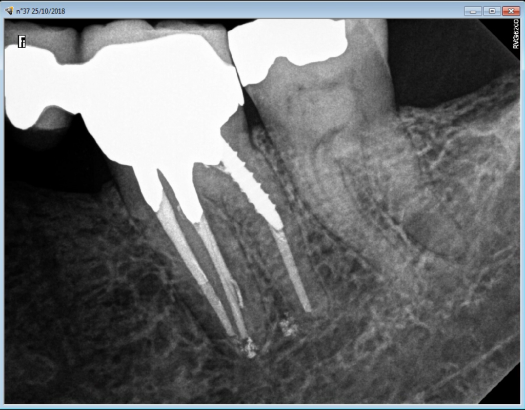
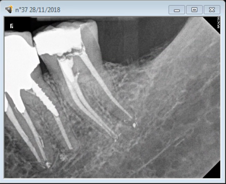
This is a unique root canal morphology were pulp chamber has two superimposed floors.

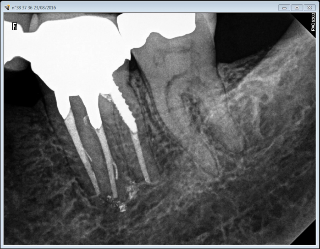
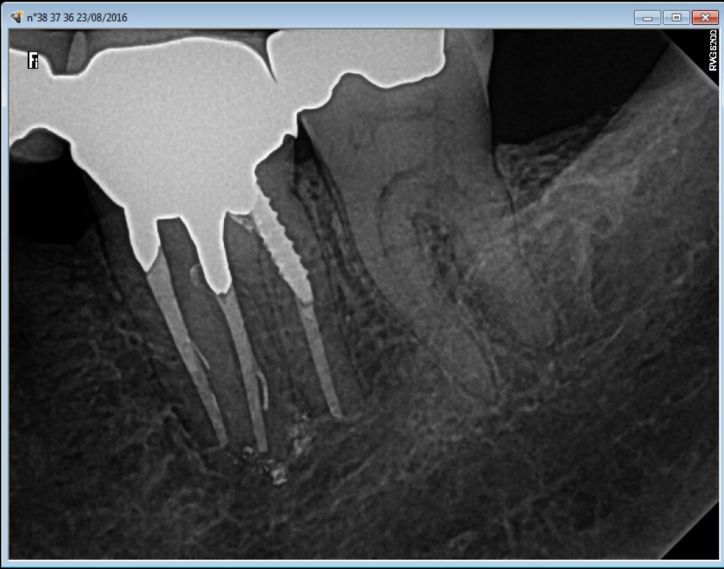
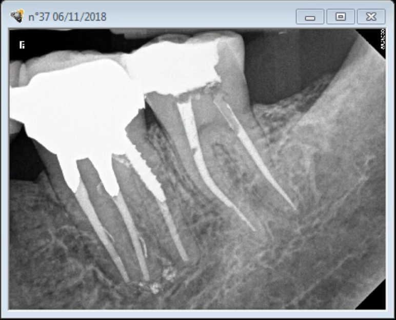
DR was analyzed with different filters. Subsequently, the access cavity was performed without the aid of DOM using Krasner and Rankow laws of symmetry and centrality. When the first (most coronal) pulp chamber floor has been reached with a 330 long shank bur the root canal entries were still obscured by reparative dentine and could not be located despite the high magnification (OPMI PRO ergo from Zeiss).

At this point, the search for the distal canal begun as it was more coronally positioned. Sclerotic dentine was thoroughly removed with a ultrasonic tip (Buc 3, Spartan) coupled to a piezo Varios 370 unit (Nakanishi) set at low power. After location of the distal root canal entry, Krasner and Rankow law of symmetry helped in locating the mesio lingual canal entriy (but not the mesio buccal one), the two canals were then negotiated and instrumented with Mani K hand files to create a glide path. Shaping completed with ProTaper Universal.

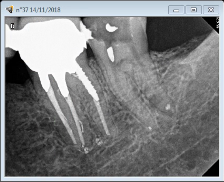
Mesio vestibular canal was much deeper and could not be located under the DOM, Krasner and Rankow laws were not sufficient since Mesio vestibular began at the deepest pulp chamber floo, thus, in order to limit the risk of perforation, patient was rescheduled as a CBCT was requested and more chair side time needed.

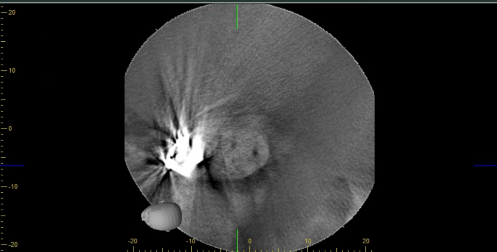
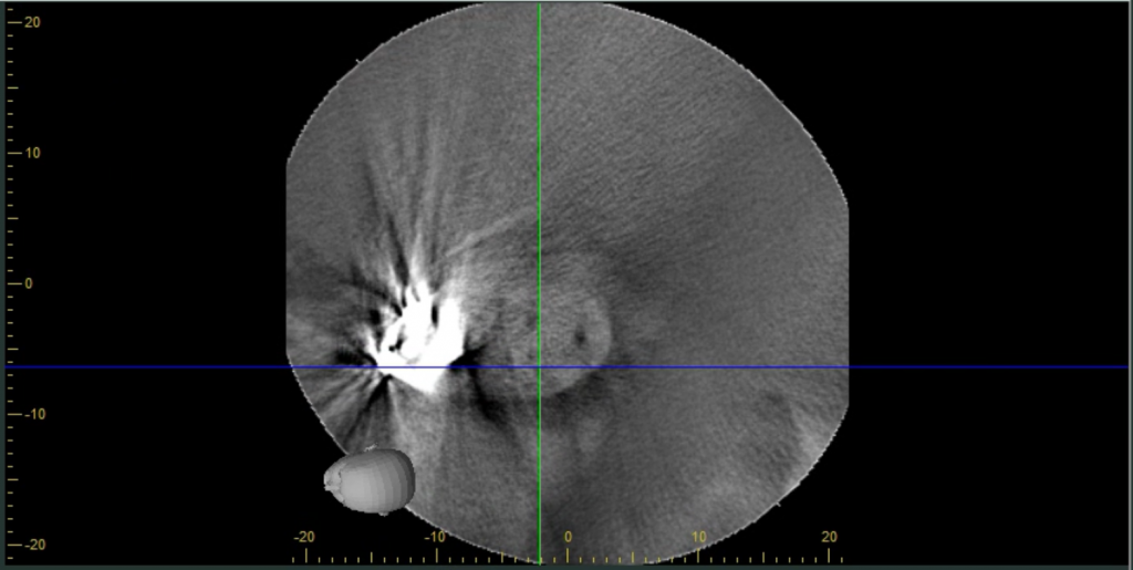
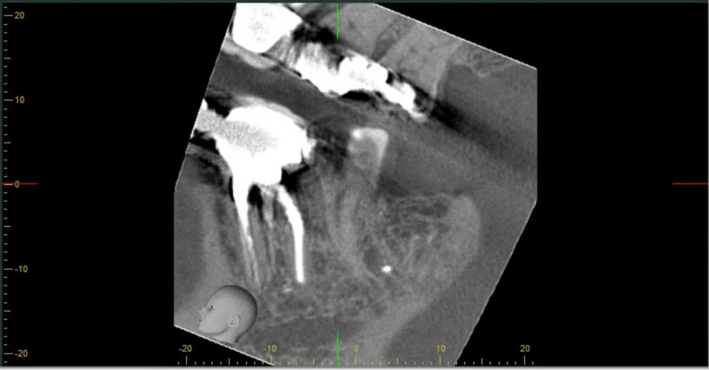
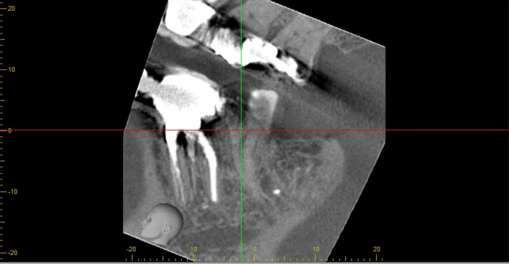
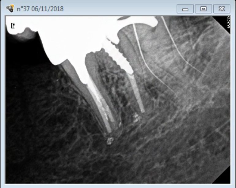
Sagittal and axial slices guided the direction of our ultrasonic tips. Mesio vestibular was finally located, an angulated X ray was taken

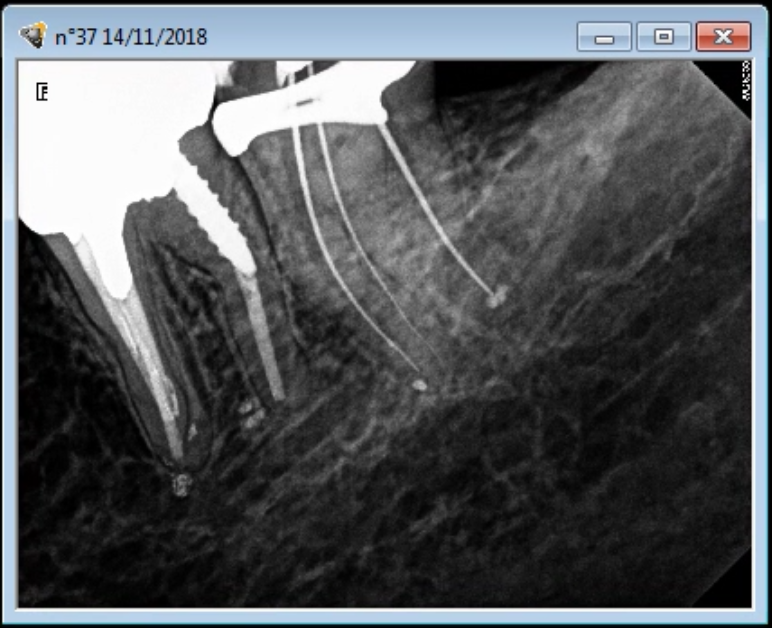
The mesio buccal canal has then been shaped, cleaned and filled with an interim Ca(OH)2 dressing.

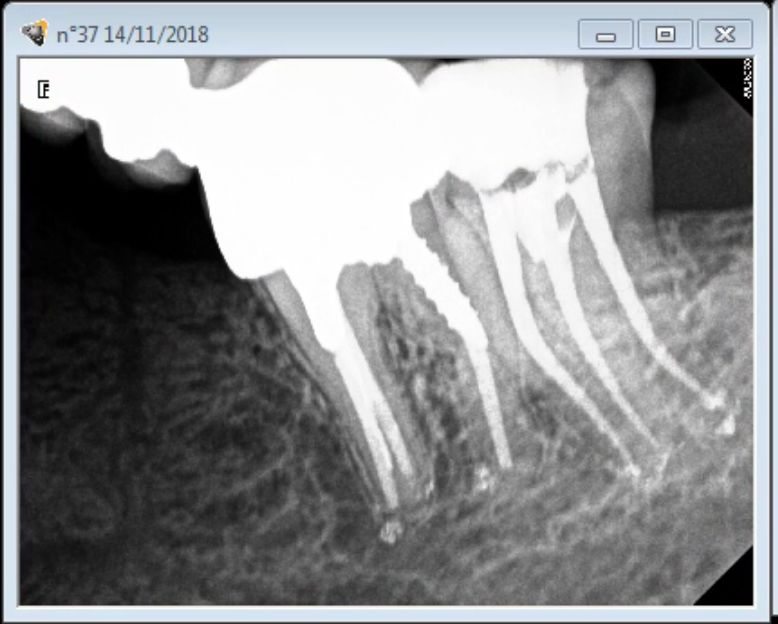
Final obturation: Canals were irrigated with 5% heated NaOCl , PUI activation. Root canal sealer used Neo MTA Plus (Avalon Biomed) cold lateral condensation.

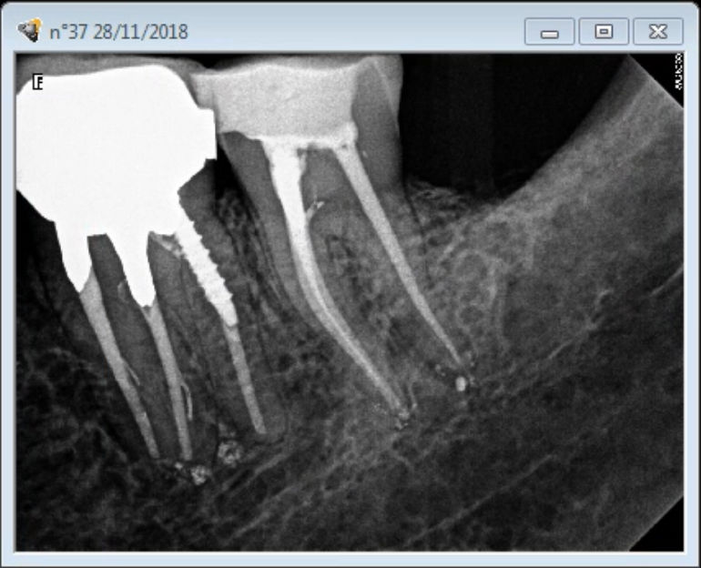
The second more apical floor could not have been cleaned by mechanical means without having to destroy a huge amount of tooth structure and without taking a risk of perforating the tooth since its filled with sclerotic dentine. Furthermore scattering was creating a lot of noise making the reading of the slices very difficult. Thus it was left alone.

Category iconCBCT assisted Root Canal Treatment,  D.O.M. versus completely calcified systems,  Dental operating microscope (D.O.M.) assisted R.C.T.,  Extreme endo clinical cases,  General,  NEW CASES,  What's new? Tag iconAvalon Biomed,  Buc 3 tips,  CBCT,  Cone-beam Computed Tomography,  dental operating microscope,  Digital Radiography,  Endodontist,  Guided Endodontics,  Krasner and Rankow,  Nakanishi,  NeoMTA Plus,  pulp canal obliteration,  pulp chamber obliteration,  root canal obliteration,  spartan,  ultrasonic tip,  Ultrasound,  Varios 370,  zeiss

Reader Interactions

Leave a Reply Cancel reply

Your email address will not be published. Required fields are marked *

Primary Sidebar

Quality Endodontics

 30+ Years of Experience
in Root Canal Treatment

 300+ Clinical Cases
Published for dentists

 Conveniently Located
Near Metro Vendôme

Call for an appointment:

(514) 667-9541

Cases with high level of difficulty:

Pierre Pizem Endodontics

A little message from Dr Pizem:

« I understand how stressful going to the dentist could be for you, the patient. So let me reassure you. I’ve built my entire reputation on my professionalism and the passion I pour into my craft. I will not compromise on equipment quality, nor on the effort necessary to restore your teeth. In the end, my greatest satisfaction is to see you fully recover your dental health, so you may grab big bites at life again. »

Ready to start your treatment?

In order to avoid risks of further complications, we recommend you book a visit as early as you can. However, if you're already feeling pain or urgent symptoms, we'll make every attempt to see you today.

Get in touch

Footer

Contact

(514) 667-9541

REQUEST APPOINTMENT

Monday
8:30 am - 4:30 pm
Tuesday
8:30 am - 4:30 pm
Wednesday
8:30 am - 4:30 pm
Thursday
8:30 am - 4:30 pm
Friday
Closed
Sat. Sun.
Closed

Clinic info

  • Home
  • About
  • Dr Pizem
  • Publications
  • Referring Dentists
    • Refer a patient
    • Clinical cases
  • Français

Patient info

  • Root Canal Therapy
  • General Dentistry
  • Endodontic Retreatment
  • FAQ New Patients
  • Tooth Saving Tips
5025 Sherbrooke St. W, Suite 330A
Montreal (QC) H4A 1S9
Near Metro Vendôme       Directions

Copyright © 2025 Endomontreal

Return to top