Patient came in our office with this very well conducted root canal (Left picture). The tooth had a radiolucid kind of filling for one year and was
Previously Underseen Second Mesiovestibular (MB2) Root Canal Location and Treatment Under Surgical Operating Microscope
Case Study Number 03
Previously Underseen Second Mesiovestibular Root Canal (MB2) Located with Surgical Operative Microscope
Case Study Number 04 Upper left first molar still symptomatic after root canal therapy. Dental operating microscope allowed us to locate second
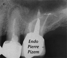
Endo Retreatment with Severe Curves Lower Left Molar
MTA Mecanical Defect Repair Above Bone Level
Case Study Number 325 I. Left side X Ray film: Iatrogenic defect 3 mm short gutta percha obturation II. Right side X Ray
MTA External Root Resorption Repair
Case Study Number 326 Pre and post MTA repair of an external resorption defect In response to a Canadian collegue concern about this MTA repair
Long Casted Post and Broken Instrument Removal to Allow for Endodontic Revision on Maxillary First Molar
Case Study Number 042210 This case shows more than one factor listed in the HIGH DIFFICULTY category: non-negotiated canal, ledge and separated
Extreme Endo to Preserve a Tooth Which May Otherwise Be Lost
Case Study Number 316 Legend: Separated file Machined post Previously underseen fourth canal apex Endo retreatment steps: Opening
Root Canal Treatment: Dealing with Casted and Machined Posts Removal
Case Study Number 409