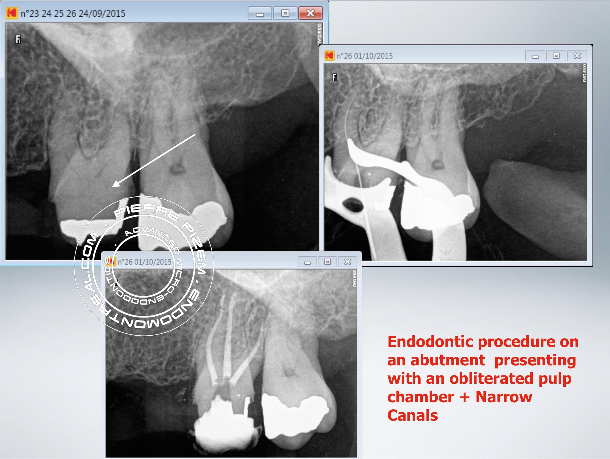
This tooth is a 23XX26 leaking fixed bridge abutment. Prior to bridge replacement it needs a root canal procedure.
Performing a root canal procedure on an abutment showing narrow canals and a complete obliteration of the pulp chamber by a large amount of calcification represents a challenge for any dentist or endodontist. Close observation of the pulpal floor map under a dental operative microscope helps in locating root canal entries. Still, this floor needs to be exposed first to be able to read the map.
Access opening was performed with a combination of a 556 cross cut and a round tungsten carbide bur (Friction Grip).
Operative field observation was enhanced with high magnification and coaxial xenon lamp illumination (Carl Zeiss OPMI PROergo dental operative microscope). Dystrophic calcifications have been removed from pulp chamber with ultrasonic diamond coated tips in order to expose the pulpal floor allowing for all canal entries location. Then, lots of chelating agent as well as many stainless steel files were needed to progressively regain patency in the narrow canals.
Instrumentation has been performed with Mani stainless K files and ProTaper Universal (Dentsply). Chelator: RC Prep. PUI Irrigation: NaOCl 5%. Ultrasonic tip: BUC One from Spartan. P5 (Dentsply) Sealer used: Pulp Canal Sealer. Obturation technique WVC. X Ray Sensors used: CareStream Kodak 6100.
Leave a Reply