• Skip to primary navigation
  • Skip to main content
  • Skip to primary sidebar
  • Skip to footer

  (514) 667-9541

  REQUEST APPOINTMENT

HOME

ABOUT

DR PIZEM

FRANÇAIS

Endomontreal Root Canal Clinic logo

ENDOMONTREAL

Dr Pizem Advanced Endodontics Clinic

  • Advanced Root Canal Treatment
    • Symptoms & Procedure
    • Why Micro Endodontics?
  • General Dentistry
  • Patients info
    • Frequently asked questions (FAQ)
    • Need a root canal revision?
    • Treatment sequence at our clinic
    • Tooth saving tips
  • For Dentists
    • Refer a patient
    • Clinical cases
    • Implants vs Endodontics
Contact Us

Huge Pulp Stone Removal Under High Magnification in Order to Preserve a Maxillary Molar

You are here: Home / Dental operating microscope (D.O.M.) assisted R.C.T. / D.O.M. versus completely calcified systems / Huge Pulp Stone Removal Under High Magnification in Order to Preserve a Maxillary Molar

May 11, 2013 By Dr Pierre Pizem Leave a Comment

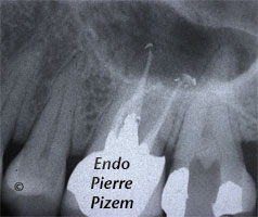
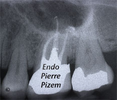
Dental operating microscope (D.O.M.), Pulp Stones (Denticles), Root Canal Treatment Pre-Therapy 479527-1Dental operating microscope (D.O.M.), Pulp Stones (Denticles), Root Canal Treatment Per-Therapy 479527-1

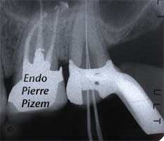
Dental operating microscope (D.O.M.), Pulp Stones (Denticles), Root Canal Treatment Per-Therapy 479527-2

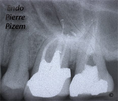
Dental operating microscope (D.O.M.), Pulp Stones (Denticles), Root Canal Treatment Per-Therapy 479527-3

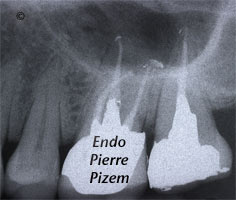
Dental operating microscope (D.O.M.), Pulp Stones (Denticles), Root Canal Treatment Post-Therapy 479527-3


Endodontic Procedure with High Magnification. Case Study Number 479527

Maxillary second left molar presenting with an acute pulpitis.

Root canal system preoperative condition: Calcified canals and a huge attached pulp stone obliterating 80% of the pulp chamber precludes any possible root canal procedure.

Procedure done under high magnification (Micro endodontics): An access cavity is made to uncover pulp stone coronal part, then a groove surrounding the attached stone is carved with a Buc 3 Ultrasonic Tip until it gets loose enough for complete removal. Unfortunately pulp stone was firmly attached to pulp chamber walls, thus, it had to be cut into pieces to allow for its removal.

Following pulp stone removal procedure, narrow calcified canals had to be located with the help of a microscope. At last, canals could then shaped, cleaned, disinfected and filled.

Category iconD.O.M. versus completely calcified systems,  Dental operating microscope (D.O.M.) assisted R.C.T.,  Extreme endo clinical cases,  General,  NEW CASES,  Popular Cases,  Pulp stones (denticles),  Root canal systems calcifications,  What's new? Tag iconcalcifications,  calcified canals,  Dystrophic calcifications,  microendodontics,  Microscope,  pulp stones,  pulpitis,  Pulpstones,  Root canal system calcifications

Reader Interactions

Leave a Reply Cancel reply

Your email address will not be published. Required fields are marked *

Primary Sidebar

Quality Endodontics

 30+ Years of Experience
in Root Canal Treatment

 300+ Clinical Cases
Published for dentists

 Conveniently Located
Near Metro Vendôme

Call for an appointment:

(514) 667-9541

Cases with high level of difficulty:

Pierre Pizem Endodontics

A little message from Dr Pizem:

« I understand how stressful going to the dentist could be for you, the patient. So let me reassure you. I’ve built my entire reputation on my professionalism and the passion I pour into my craft. I will not compromise on equipment quality, nor on the effort necessary to restore your teeth. In the end, my greatest satisfaction is to see you fully recover your dental health, so you may grab big bites at life again. »

Ready to start your treatment?

In order to avoid risks of further complications, we recommend you book a visit as early as you can. However, if you're already feeling pain or urgent symptoms, we'll make every attempt to see you today.

Get in touch

Footer

Contact

(514) 667-9541

REQUEST APPOINTMENT

Monday
8:30 am - 4:30 pm
Tuesday
8:30 am - 4:30 pm
Wednesday
8:30 am - 4:30 pm
Thursday
8:30 am - 4:30 pm
Friday
Closed
Sat. Sun.
Closed

Clinic info

  • Home
  • About
  • Dr Pizem
  • Publications
  • Referring Dentists
    • Refer a patient
    • Clinical cases
  • Français

Patient info

  • Root Canal Therapy
  • General Dentistry
  • Endodontic Retreatment
  • FAQ New Patients
  • Tooth Saving Tips
5025 Sherbrooke St. W, Suite 330A
Montreal (QC) H4A 1S9
Near Metro Vendôme       Directions

Copyright © 2025 Endomontreal

Return to top