Case Study Number 486726 Patient was referred to us with a partial pulpectomy in 3 out of four canals. The case came with a
Locating MB2 and DB Orifices with Dental Operative Microscope on Calcified Canals in a Maxillary First Molar
Case Study Number 434426 Patient was refered to us to locate MB2 and DB canal entries with the help of a dental operative microscope.
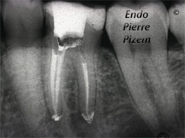
Mesial Calcified Canals Entries Location Under High Magnification
An intricate root canal procedure, because this pre operative condition involves dealing with complete canal stenosis caused by dystrophic
Dealing with a Crowned Maxillary Molar and Extreme Calcifications in it’s Root Canal System
Endodontic Procedure Under Dental Operative Microscope. Case Study Number 282816 Surgical operating microscope assisted root canal treatment through
Fiberglass Reinforced Composite Posts Removal in Both Maxillary Premolar to Allow Endodontic Retreatment
Root Canal Procedure. Case Study Number 1 First premolar was having an under fill in its distovestibular root canal. Meaning that one small diameter
Calcified Canals and Root Canal Treatment
Root Canal Procedure on Calcified Canals. Endodontic Microscope. Case Study Number 414 Second mesiobuccal canal entry (MB-2) in this upper molar was
Extreme Endo on Calcified Canals to Preserve Two Teeth which May Otherwise Be Lost
Root Canal Procedure on Calcified Canals. Endodontic. Case Study Number 102334 The patient was