• Skip to primary navigation
  • Skip to main content
  • Skip to primary sidebar
  • Skip to footer

  (514) 667-9541

  REQUEST APPOINTMENT

HOME

ABOUT

DR PIZEM

FRANÇAIS

Endomontreal Root Canal Clinic logo

ENDOMONTREAL

Dr Pizem Advanced Endodontics Clinic

  • Advanced Root Canal Treatment
    • Symptoms & Procedure
    • Why Micro Endodontics?
  • General Dentistry
  • Patients info
    • Frequently asked questions (FAQ)
    • Need a root canal revision?
    • Treatment sequence at our clinic
    • Tooth saving tips
  • For Dentists
    • Refer a patient
    • Clinical cases
    • Implants vs Endodontics
Contact Us

Images

You are here: Home / Archives for Images

February 16, 2012

Microendodontics with Carl Zeiss OPMI PROergo Dental Operative Microscope. Root Canal Treatment Procedure on a Second Maxillary Molar with Calcified Canals

A new Dental Operative Microscope (D.O.M.) assisted root canal treatment on a maxillary second molar with calcified canals. MicroEndodontics. Case

Category iconD.O.M. versus completely calcified systems,  Dental operating microscope (D.O.M.) assisted R.C.T.,  Root canal systems calcifications,  Striving for second Mesio vestibular (MB2),  What's new? Tag iconAdherent pulpstones,  calcified canals,  calcified dentine,  complete root canal stenosis,  Dental operative microscope,  dentin,  dystrophic,  endodontic files,  Endodontics,  Endodontist,  garry carr,  Gutta percha,  Images,  microendodontics,  Opmi Proergo microscope,  pro taper,  Protaper dentsply,  pulp canal sealer,  root,  Root canal,  root canal perforation,  root canal sealer,  Root canal system calcifications

December 2, 2011

To Save or Not To Save? That Was the Question. A Seven Years Post Endodontic Treatment Outcome Follow Up

       Case Study Number  368745 Patient was told seven years ago to remove lower right premolar and replace this tooth by an implant supported

Category iconDental operative microscope and retreatment,  Extreme endo clinical cases,  NEW CASES,  Post endodontic treatment outcomes,  Regeneration of the periradicular tissues,  What's new? Tag iconEndodontic expertise,  endodontic procedure,  Endodontics,  Endodontist,  follow up,  Gutta percha,  Images,  microendodontics,  pro taper,  pulp canal sealer,  regeneration of periradicular tissues,  Retreatment,  Root canal,  Root canal treatment

November 17, 2011

Microendodontics with Carl Zeiss OPMI PROergo Dental Operative Microscope. Root Canal Treatment Procedure on a Lateral Incisor with a Calcified Canal

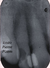
                                     A new Dental Operative Microscope (D.O.M.) Assisted Root Canal Treatment in a Calcified Maxillary Lateral

Category iconD.O.M. versus completely calcified systems,  Dental operating microscope (D.O.M.) assisted R.C.T.,  Extreme endo clinical cases,  NEW CASES,  What's new? Tag iconAdherent pulpstones,  calcified canal,  calcified dentine,  complete root canal stenosis,  Dental operative microscope,  dentin,  dystrophic,  endodontic files,  Endodontics,  Endodontist,  garry carr,  Gutta percha,  Images,  microendodontics,  Opmi Proergo microscope,  pro taper,  Protaper dentsply,  pulp canal sealer,  root,  Root canal,  root canal perforation,  root canal sealer,  Root canal system calcifications

October 13, 2011

An Intricate Root Canal Procedure on a Mineralized Second Maxillary Molar with a Canal Curvature into an “S” Form

    Endodontic Procedure. Case Study Number 449927 To treat such a tooth in endodontics we needed to deal with: Difficult access Long tooth

Category iconAtypical canal configurations,  Canal curvature with an 'S' form,  Curved canals,  D.O.M. versus partially calcified systems,  Dental operating microscope (D.O.M.) assisted R.C.T.,  Extreme endo clinical cases,  NEW CASES,  Striving for second Mesio vestibular (MB2) Tag iconbayonet shaped root canal,  canal curvature with an s form,  Dental expertise,  Dental operative microscope,  Denticles,  Dystrophic calcifications,  Endodontic expertise,  endodontic procedure,  Endodontics,  Endodontist,  Gutta percha,  Images,  Location,  Mb2,  Mesiovestibular,  microendodontics,  Opmi Proergo microscope,  Protaper dentsply,  pulp canal sealer,  Root canal,  root canal procedure,  Root canal system calcifications,  Root canal treatment

October 7, 2011

An Intricate Root Canal Procedure on a Severely Curved Root Canal System with Pulp Tissue Fibrosis

    MicroEndodontic. Case Study Number 500047 Canal curvatures are a challenge to preparation and can be the origin of many technical complications

Category iconCurved canals,  D.O.M. versus partially calcified systems,  Dental operating microscope (D.O.M.) assisted R.C.T.,  Extreme endo clinical cases,  Extremely curved root canals (90 degrees +),  NEW CASES,  Pulp stones (denticles) Tag iconDental expertise,  Dental operative microscope,  endodontic microscope,  Endodontist,  Gutta percha,  Images,  microendodontics,  Opmi Proergo microscope,  pro taper,  pulp canal sealer,  root canal procedure

September 14, 2011

Microendodontics with CARL ZEISS OPMI PROergo VS Completely Calcified Canal

        A New Dental Operative Microscope (D.O.M.) Assisted Root Canal Treatment on Maxillary Incisor with a Calcified Canal. MicroEndodontic. Case

Category iconD.O.M. versus completely calcified systems,  Dental operating microscope (D.O.M.) assisted R.C.T.,  Extreme endo clinical cases,  NEW CASES Tag iconcalcified canal,  calcified dentine,  Calcium hydroxide,  canal stenosis,  Carl Zeiss,  complete root canal stenosis,  Dental operative microscope,  Dystrophic calcifications,  Endodontist,  Gutta percha,  Images,  intricate,  Location,  microendodontics,  pulp canal sealer,  radiolucent apical lesion,  root canal perforation,  root canal procedure,  Root canal system calcifications,  Root canal treatment,  surgical operative microscope,  treatment protocol

September 8, 2011

Radix Entomolaris Presenting a Fifth Canal (Accessory Canal) Extending from the Pulp Chamber to the Furcation

        Microendodontic. Case Study Number 493847 Key words: Root canal anatomy, anatomical variation of teeth, radix entomolaris The anatomy of

Category iconAtypical canal configurations,  Dental operating microscope (D.O.M.) assisted R.C.T.,  Extreme endo clinical cases,  NEW CASES,  Radix Entomolaris Tag iconanatomical variation of teeth,  Calcium hydroxide,  Coronal-radicular access,  Dental operative microscope,  endodontic procedure,  Endodontist,  Gutta percha,  Images,  Location,  microendodontics,  Opmi Proergo microscope,  pulp canal sealer,  radiolucent apical lesion,  Root canal,  root canal anatomy,  Root canal treatment

September 1, 2011

OPMI PROergo Dental Operative Microscope VS Extreme Root Canal System Stenosis on a Maxillary Molar

      A New Dental Operative Microscope (D.O.M.) Assisted Root Canal Treatment in a Calcified Maxillary Molar Abutment. Microendodontic. Case Study

Category iconD.O.M. versus completely calcified systems,  Dental operating microscope (D.O.M.) assisted R.C.T.,  Extreme endo clinical cases,  NEW CASES,  Root canal systems calcifications Tag iconAdherent pulpstones,  calcified canals,  calcified dentine,  complete root canal stenosis,  Dental operative microscope,  dentin,  dentin map,  dentin maps,  Dystrophic calcifications,  endodontic files,  Endodontics,  Endodontist,  gary carr,  Gutta percha,  Images,  microendodontics,  Opmi Proergo microscope,  pro taper,  Protaper dentsply,  pulp canal sealer,  Root canal,  root canal perforation,  root canal procedure,  Root canal system calcifications

August 27, 2011

Metalift Crown and Bridge Removal System to save an existing bridge. Another success story.

  Case Study Number 497935 Patient has already been treated on an emergency basis for an acute apical periodontitis with an irreversible

Category iconCanal curvature with an 'S' form,  Curved canals,  Dental operating microscope (D.O.M.) assisted R.C.T.,  NEW CASES,  Popular Cases,  Pulp stones (denticles),  Root canal systems calcifications Tag iconDental operative microscope,  Denticles,  Dystrophic calcifications,  endodontic procedure,  Endodontics,  Endodontist,  Gutta percha,  Images,  Location,  metalift,  microendodontics,  Microscope,  Opmi Proergo microscope,  pulp canal sealer,  Pulpstones,  root canal procedure,  Root canal system calcifications,  Root canal treatment

  • Go to page 1
  • Go to page 2
  • Go to page 3
  • Go to page 4
  • Go to Next Page »

Primary Sidebar

Quality Endodontics

 25+ Years of Experience
in Root Canal Treatment

 300+ Clinical Cases
Published for dentists

 Conveniently Located
Near Metro Vendôme

Call for an appointment:

(514) 667-9541

Cases with high level of difficulty:

Pierre Pizem Endodontics

A little message from Dr Pizem:

« I understand how stressful going to the dentist could be for you, the patient. So let me reassure you. I’ve built my entire reputation on my professionalism and the passion I pour into my craft. I will not compromise on equipment quality, nor on the effort necessary to restore your teeth. In the end, my greatest satisfaction is to see you fully recover your dental health, so you may grab big bites at life again. »

Ready to start your treatment?

In order to avoid risks of further complications, we recommend you book a visit as early as you can. However, if you're already feeling pain or urgent symptoms, we'll make every attempt to see you today.

Get in touch

Footer

Contact

(514) 667-9541

REQUEST APPOINTMENT

Monday
8:30 am - 4:30 pm
Tuesday
8:30 am - 4:30 pm
Wednesday
8:30 am - 4:30 pm
Thursday
8:30 am - 4:30 pm
Friday
8:30 am - 4:30 pm
Sat. Sun.
Closed

Clinic info

  • Home
  • About
  • Dr Pizem
  • Publications
  • Referring Dentists
    • Refer a patient
    • Clinical cases
  • Français

Patient info

  • Root Canal Therapy
  • General Dentistry
  • Endodontic Retreatment
  • FAQ New Patients
  • Tooth Saving Tips
5025 Sherbrooke St. W, Suite 330A
Montreal (QC) H4A 1S9
Near Metro Vendôme       Directions

Copyright © 2023 Endomontreal

Return to top