• Skip to primary navigation
  • Skip to main content
  • Skip to primary sidebar
  • Skip to footer

  (514) 667-9541

  REQUEST APPOINTMENT

HOME

ABOUT

DR PIZEM

FRANÇAIS

Endomontreal Root Canal Clinic logo

ENDOMONTREAL

Dr Pizem Advanced Endodontics Clinic

  • Advanced Root Canal Treatment
    • Symptoms & Procedure
    • Why Micro Endodontics?
  • General Dentistry
  • Patients info
    • Frequently asked questions (FAQ)
    • Need a root canal revision?
    • Treatment sequence at our clinic
    • Tooth saving tips
  • For Dentists
    • Refer a patient
    • Clinical cases
    • Implants vs Endodontics
Contact Us

Gutta percha

You are here: Home / Archives for Gutta percha
calcified-curved-and-long-canals-26c-2016-12-03 Post therapy A

December 3, 2016

A Root Canal on a Calcified, Curvy Long Tooth

   RCT and amalgam core build up on a very long, remote and heavily calcified 26 (Irreversible Pulpitis) Endodontic procedure instrumentation has

Category iconD.O.M. versus partially calcified systems,  Dental operating microscope (D.O.M.) assisted R.C.T.,  Extreme endo clinical cases,  Extremely curved root canals (90 degrees +),  Post and core build ups,  Pulp stones (denticles),  Root canal systems calcifications,  Very long teeth Tag iconanatomical variation,  Gutta percha,  mani endodontic files,  Opmi Proergo microscope,  protaper universal,  pulp canal sealer,  rc prep,  root canal calcifications,  root canal curvature,  surgical operative microscope,  very long tooth,  zeiss

Performing root canal revision procedure

June 27, 2013

Uncovered Vertucci’s Type V in Distal Root During Endodontic Revision 7 Years Ago Allowed for that Tooth to be Preserved. Thanks to OPMI PROergo Dental Operative Microscope

Dental operative microscope root canal procedure (Microendodontics). Case Study Number 392346. Referred patient has had two root canal

Category iconAtypical canal configurations,  Dental operating microscope (D.O.M.) assisted R.C.T.,  Dental operative microscope and retreatment,  Extreme endo clinical cases,  General,  NEW CASES,  Type V,  What's new? Tag iconcalcium hydroxyde,  dental implant,  dental implant versus natural tooth,  Dental operative microscope,  difficult,  endodontic microscope,  endodontic revision,  Endodontist,  Gutta percha,  high level of difficulty,  microendodontics,  Opmi Proergo microscope,  Pain,  pain relief,  pulp canal sealer,  root canal procedure,  vertucci type V root canal configuration

Post removal and endodontic revision

June 7, 2013

This Patient Chose Orthograde Endodontic Revision Procedure on his Mandibular Premolar Instead of Extraction and a Dental Implant

Because of the huge post cemented within its root canal, the endodontic revision procedure to save her tooth seemed to be too difficult or too risky

Category iconD.O.M. versus partially calcified systems,  Dealing with casted and machined posts removal,  Dealing with ledges and apical zip,  Dealing with N2 (Sargenti Paste) removal,  Dental operating microscope (D.O.M.) assisted R.C.T.,  Dental operative microscope and retreatment,  NEW CASES,  Root canal systems calcifications,  What's new? Tag iconbone destruction,  calcified canal,  casted post,  condensation technique,  endodontic microscope,  endodontic revision,  Endodontist,  Gutta percha,  ledge,  Microscope,  Opmi Proergo microscope,  pulp canal sealer root canal procedure,  sargenti paste

June 21, 2012

Root Canal Procedure on Radix Entomolaris Presenting a Severe Dilaceration

Microscope in Endododontics. Case Study Number 513236. We have been confronted to this three rooted mandibular first molar (Radix Molar or Radix

Category iconAtypical canal configurations,  Canal curvature with an 'S' form,  Curved canals,  D.O.M. versus partially calcified systems,  Dental operating microscope (D.O.M.) assisted R.C.T.,  Dilaceration,  Extremely curved root canals (90 degrees +),  Radix Entomolaris Tag iconcalcifications,  condensation technique,  dilacerated curve,  dilacerated teeth,  dilacerated tooth,  dilaceration,  Donald Yu,  endodontic file,  endodontic microscope,  endodontic procedure,  Endodontist,  endomontreal,  Gutta percha,  Protaper dentsply,  pulp canal sealer,  Pulpstones,  radius of curvature,  radix entomolaris,  radix molar,  root canal procedure,  Root canal system calcifications,  schilder,  small radius

April 13, 2012

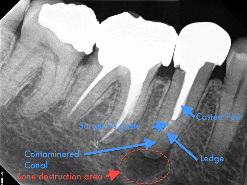
A Root Canal Retreatment Involving a Crown and Two Posts Removal allowed for this Painful Tooth to Be Preserved

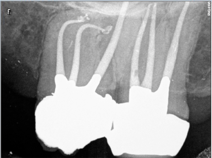
    Microendodontic. Case Study Number 511536 Sixty years old patient presenting with an abscessed mandibular molar. Diagnosis: Persisting disease

Category iconDealing with casted and machined posts removal,  Dealing with ledges and apical zip,  Dental operative microscope and retreatment,  What's new? Tag iconabscess,  case report,  clip,  crown,  dental implant,  endodontic retreatment,  extraction,  farzaneh,  Gutta percha,  microendodontics,  persisting disease,  post,  post removal,  prognosis,  pulp canal sealer,  Retreatment,  root canal filling,  root canal system,  Root canal treatment,  save,  tooth,  voco

February 16, 2012

Microendodontics with Carl Zeiss OPMI PROergo Dental Operative Microscope. Root Canal Treatment Procedure on a Second Maxillary Molar with Calcified Canals

A new Dental Operative Microscope (D.O.M.) assisted root canal treatment on a maxillary second molar with calcified canals. MicroEndodontics. Case

Category iconD.O.M. versus completely calcified systems,  Dental operating microscope (D.O.M.) assisted R.C.T.,  Root canal systems calcifications,  Striving for second Mesio vestibular (MB2),  What's new? Tag iconAdherent pulpstones,  calcified canals,  calcified dentine,  complete root canal stenosis,  Dental operative microscope,  dentin,  dystrophic,  endodontic files,  Endodontics,  Endodontist,  garry carr,  Gutta percha,  Images,  microendodontics,  Opmi Proergo microscope,  pro taper,  Protaper dentsply,  pulp canal sealer,  root,  Root canal,  root canal perforation,  root canal sealer,  Root canal system calcifications

February 10, 2012

Endodontic Revision Procedure on Mandibular Molar, a 6 months Post Operative Outcome

Case Study Number 495336 Symptoms: Acute pain to pressure, patient is eating on the opposite side. Root canal was done three years ago. Tooth

Category iconDealing with N2 (Sargenti Paste) removal,  Dental operative microscope and retreatment,  Post endodontic treatment outcomes,  Regeneration of the periradicular tissues,  What's new? Tag iconendodontic files,  endodontic microscope,  endodontic revision,  Gutta percha,  outcomes,  pulp canal sealer,  root canal procedure,  root canal system,  Root canal treatment

January 19, 2012

Endodontic Treatment on Mandibular Molar with Calcified Canals and a “J” Type Lesion, a Five Years Follow Up

       Root Canal Procedure with Surgical Operative Microscope. MicroEndodontic. Case Study Number 156037. Pre operative film shows a large bony

Category iconD.O.M. versus completely calcified systems,  Dental operating microscope (D.O.M.) assisted R.C.T.,  Extreme endo clinical cases,  NEW CASES,  Popular Cases,  Post endodontic treatment outcomes,  Pulp stones (denticles),  Regeneration of the periradicular tissues,  Root canal systems calcifications,  What's new? Tag iconAdherent pulpstones,  calcified canals,  complete root canal stenosis,  Embedded pulpstones,  endodontic files,  Gutta percha,  intracanal medication,  rc prep,  Root canal,  root canal procedure,  root canal sealer,  root canal system,  Root canal system calcifications,  Root canal treatment,  sodium hypochlorite,  treatment protocol

December 2, 2011

To Save or Not To Save? That Was the Question. A Seven Years Post Endodontic Treatment Outcome Follow Up

       Case Study Number  368745 Patient was told seven years ago to remove lower right premolar and replace this tooth by an implant supported

Category iconDental operative microscope and retreatment,  Extreme endo clinical cases,  NEW CASES,  Post endodontic treatment outcomes,  Regeneration of the periradicular tissues,  What's new? Tag iconEndodontic expertise,  endodontic procedure,  Endodontics,  Endodontist,  follow up,  Gutta percha,  Images,  microendodontics,  pro taper,  pulp canal sealer,  regeneration of periradicular tissues,  Retreatment,  Root canal,  Root canal treatment

  • Go to page 1
  • Go to page 2
  • Go to page 3
  • Go to Next Page »

Primary Sidebar

Quality Endodontics

 25+ Years of Experience
in Root Canal Treatment

 300+ Clinical Cases
Published for dentists

 Conveniently Located
Near Metro Vendôme

Call for an appointment:

(514) 667-9541

Cases with high level of difficulty:

Pierre Pizem Endodontics

A little message from Dr Pizem:

« I understand how stressful going to the dentist could be for you, the patient. So let me reassure you. I’ve built my entire reputation on my professionalism and the passion I pour into my craft. I will not compromise on equipment quality, nor on the effort necessary to restore your teeth. In the end, my greatest satisfaction is to see you fully recover your dental health, so you may grab big bites at life again. »

Ready to start your treatment?

In order to avoid risks of further complications, we recommend you book a visit as early as you can. However, if you're already feeling pain or urgent symptoms, we'll make every attempt to see you today.

Get in touch

Footer

Contact

(514) 667-9541

REQUEST APPOINTMENT

Monday
8:30 am - 4:30 pm
Tuesday
8:30 am - 4:30 pm
Wednesday
8:30 am - 4:30 pm
Thursday
8:30 am - 4:30 pm
Friday
8:30 am - 4:30 pm
Sat. Sun.
Closed

Clinic info

  • Home
  • About
  • Dr Pizem
  • Publications
  • Referring Dentists
    • Refer a patient
    • Clinical cases
  • Français

Patient info

  • Root Canal Therapy
  • General Dentistry
  • Endodontic Retreatment
  • FAQ New Patients
  • Tooth Saving Tips
5025 Sherbrooke St. W, Suite 330A
Montreal (QC) H4A 1S9
Near Metro Vendôme       Directions

Copyright © 2023 Endomontreal

Return to top