This is a symptomatic mandibular first premolar (bridge abutment) with a "C" shape root canal system. Most C-shaped canals occurs in mandibular second
Mesial Calcified Canals Entries Location Under High Magnification
An intricate root canal procedure, because this pre operative condition involves dealing with complete canal stenosis caused by dystrophic
Endodontic Dentistry: Double “C” Shape Distal Canal VS Carl Zeiss OPMI Dental Operative Microscope
Case Study Number 331737 Heated gutta percha was most useful to obtain that so sought after 3D obturation in this mandibular second molar. En esta
First Maxillary Molar with Complete Calcification of its Canal System Preserved with a Microscope Assisted Root Canal (Case 471726)
Patient is experiencing severe pain on upper left side and is seen on an emergency basis. The first molar is having a necrotic pulp and an acute
Root Canal Procedure on Calcified Canals, Striving for Second Mesio Vestibular Root Canal Treatment
Root Canal Procedure on Calcified Canals. Case Study Number 413
Broken NITI File Retrieval in Mesiovestibular Root of a Maxillary Molar
Case Study Number 264266 Tooth presenting an " against all odds " clinical situation , many would think " you don't have to worry it is dead! "
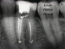
Endodontic Treatment on Mandibular Molar with Complete Stenosis of Mesial Root Canals (Calcified Canals)
Calcified Canals. Root Canal Procedure. Case Study Number 119036 Pre operative film shows a large bony defect reminding us the
Dental Operative Microscope Assisted Endodontic Treatment of Sclerotic Canals (Calcified Canals)
Root Canal Procedure on Calcified Canals. Case Study Number 91724 Symptomatic first maxillary premolar with deciduous extensive composite restoration
Root Canal and Pulp Chamber Not Visible on Pre Operative X Ray of Maxillary Lateral Incisor (Calcified Canals)
Root Canal Procedure on Calcified Canals. Case Study Number 219222