• Skip to primary navigation
  • Skip to main content
  • Skip to primary sidebar
  • Skip to footer

  (514) 667-9541

  REQUEST APPOINTMENT

HOME

ABOUT

DR PIZEM

FRANÇAIS

Endomontreal Root Canal Clinic logo

ENDOMONTREAL

Dr Pizem Advanced Endodontics Clinic

  • Advanced Root Canal Treatment
    • Symptoms & Procedure
    • Why Micro Endodontics?
  • General Dentistry
  • Patients info
    • Frequently asked questions (FAQ)
    • Need a root canal revision?
    • Treatment sequence at our clinic
    • Tooth saving tips
  • For Dentists
    • Refer a patient
    • Clinical cases
    • Implants vs Endodontics
Contact Us

Dental operating microscope (D.O.M.) assisted R.C.T.

You are here: Home / Archives for Dental operating microscope (D.O.M.) assisted R.C.T.
2MB root canal 2DB root canal1P root canal Post operative 2016-03-08

March 14, 2016

Treating a maxillary molar with 5 canals and 5 portals of exit

Anatomical variations must be considered at the beginning of the root canal treatment. Even though at this point, accurate preoperative radiographs

Category iconAtypical canal configurations,  Dental operating microscope (D.O.M.) assisted R.C.T.,  Extreme endo clinical cases,  NEW CASES,  Pulp stones (denticles) Tag iconanatomical variation,  magnification,  rare,  Root canal,  Root canal treatment

Root canal procedure on a maxillary molar presenting with a supernumerary palatal root

March 10, 2016

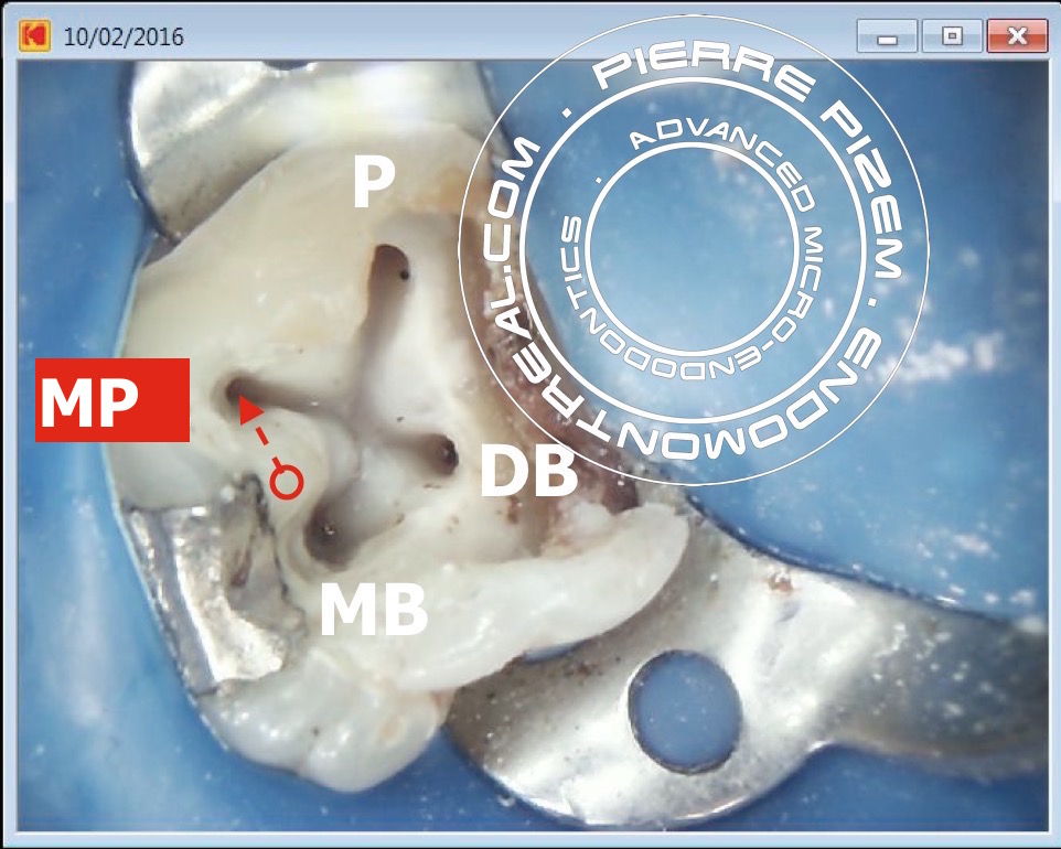
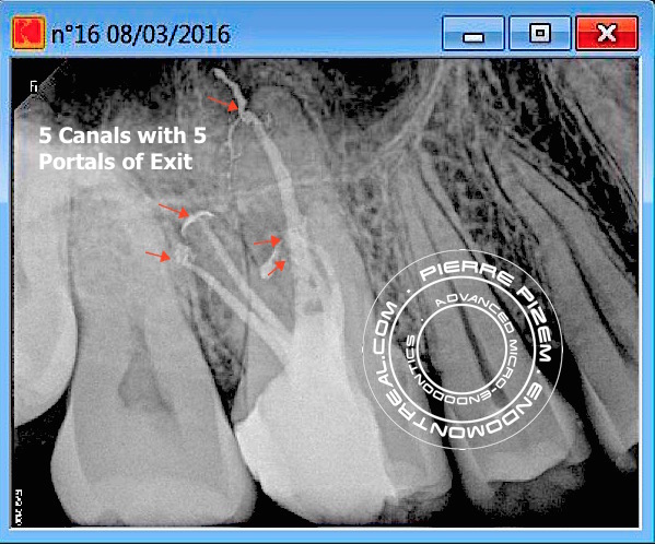
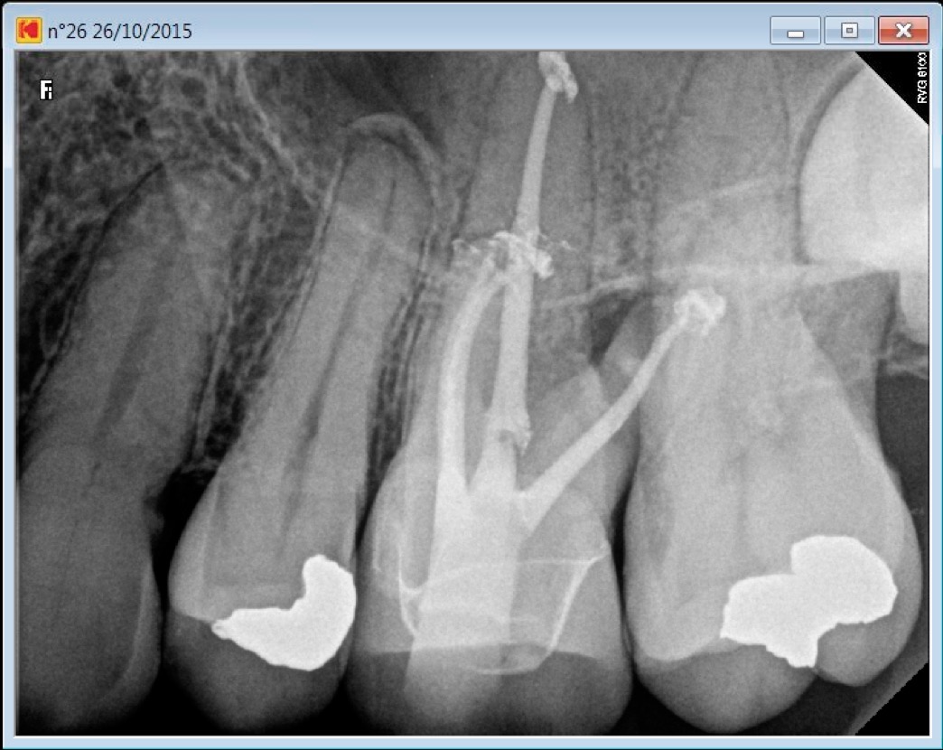
A root canal procedure on a molar presenting with a supernumerary mesio palatal (MP) root

         Root canal procedure on molar presenting with an extra mesio palatal (MP) root. A very rare anatomical variation. This supernumerary

Category iconAtypical canal configurations,  Dental operating microscope (D.O.M.) assisted R.C.T.,  General,  NEW CASES,  Radix Entomolaris Tag iconaccess cavity,  carestream kodak 6100,  endodontic instruments,  endodontic procedure,  mani stainless endodontic files,  protaper universal,  pulp canal sealer,  pulp chamber,  Root canal,  root canal procedure,  shaping,  sodium hypochlorite,  supernumerary root,  warm vertical condensation

46 pre therapy 2016-02-09

February 20, 2016

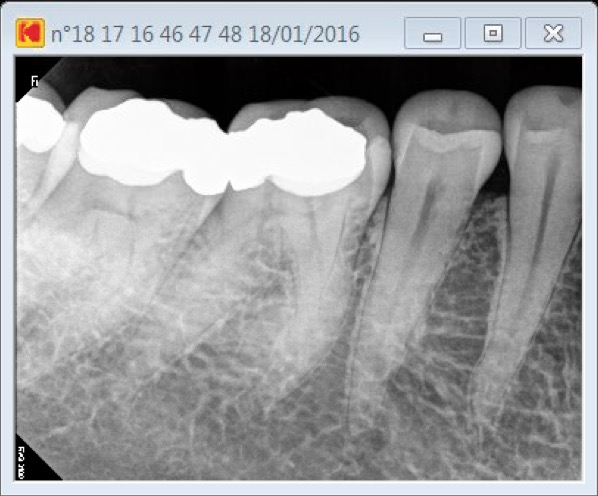
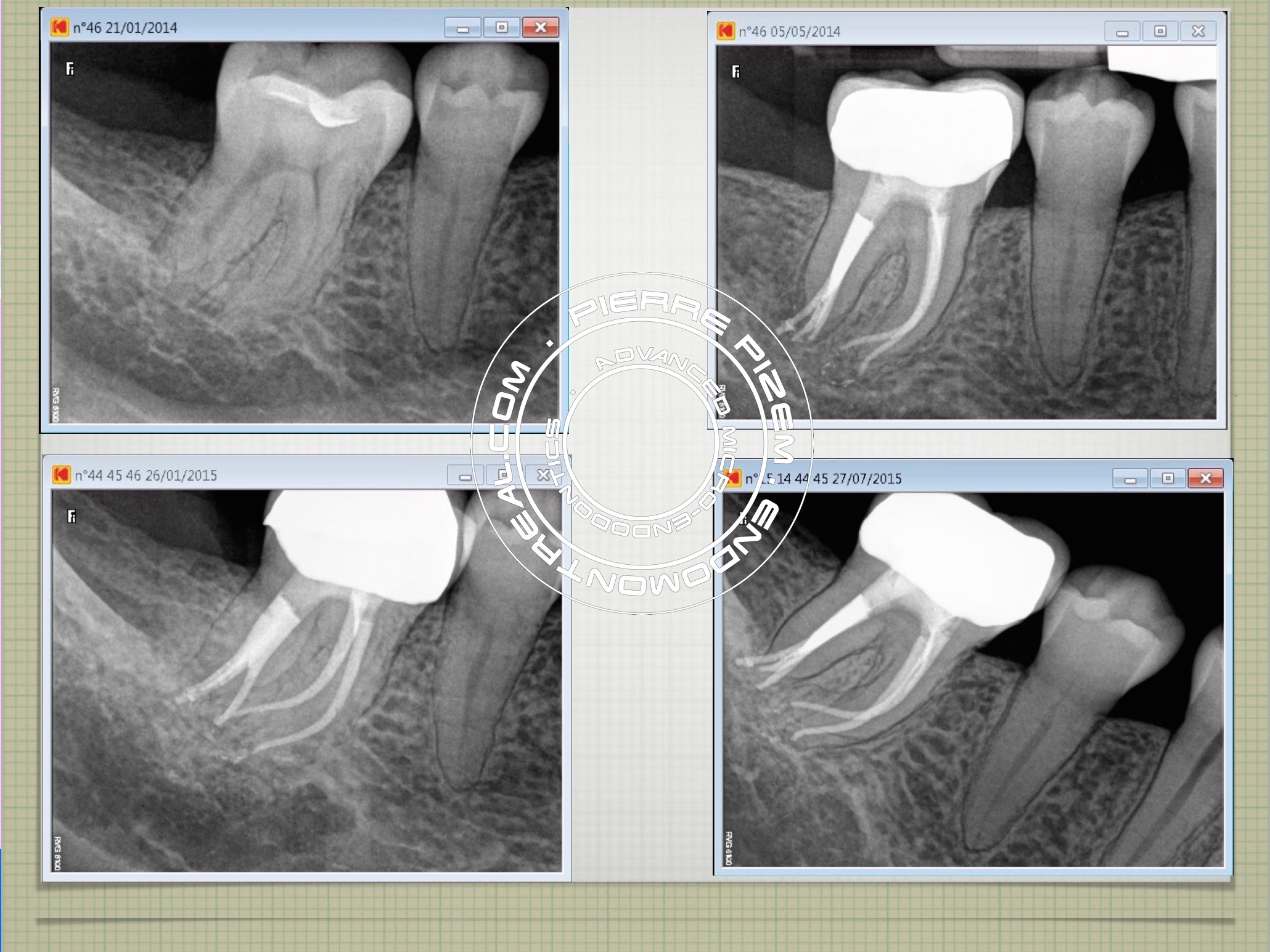
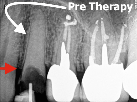
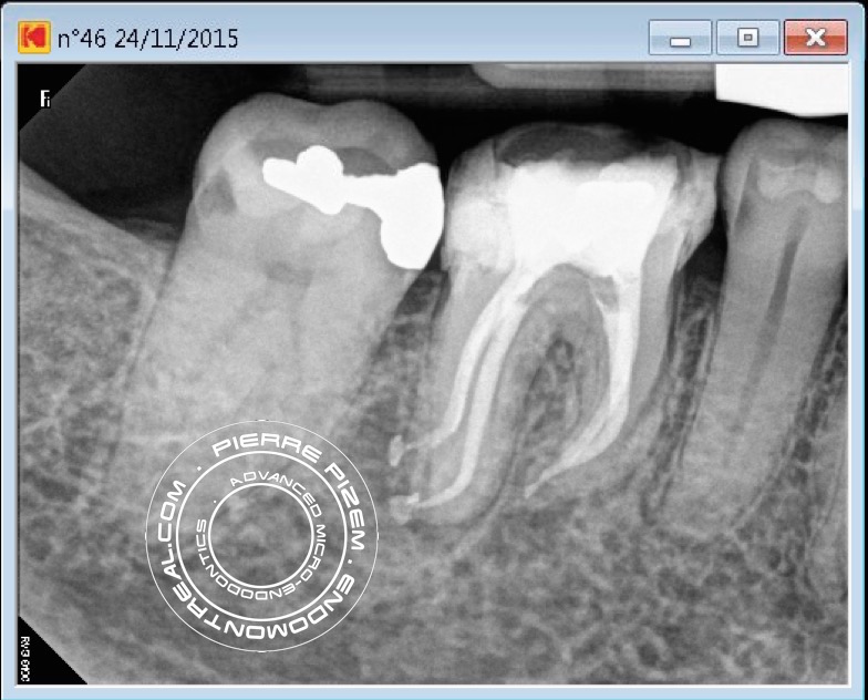
A root canal treatment on a calcified molar

   Root canal system is barely visible on pre operative dental X ray image indicating almost complete pulp chamber and root canal stenosis. Root

Category iconD.O.M. versus partially calcified systems,  Dental operating microscope (D.O.M.) assisted R.C.T.,  Extreme endo clinical cases,  Pulp stones (denticles) Tag iconchelator,  complete root canal stenosis,  endodontic microscope,  mani endodontic files,  nayyar,  rc prep,  root canal calcifications,  root canal entries,  Root canal treatment

One canal and three portals of exit in distal root 46

February 5, 2016

An Endodontic Procedure on a Unique Root Canal Morphology

This case has been posted on a specialized root canal FB forum and earned 596 likes from dentists around the world. See this case at:  International

Category iconAtypical canal configurations,  Canal curvature with an 'S' form,  Dental operating microscope (D.O.M.) assisted R.C.T. Tag icondentsply,  protaper universal,  root canal anatomy,  root canal apical split,  root canal morphology,  root canal procedure,  root canal system

January 8, 2016

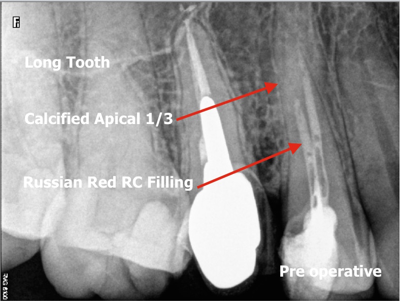
A natural tooth merely escaping from extraction

   Due to the presence of rock hard Russian Red filling material in its root canal, the endodontic revision procedure to save her tooth seemed to

Category iconD.O.M. versus completely calcified systems,  Dealing with N2 (Sargenti Paste) removal,  General Tag iconblockage,  chelating agent,  dental implant,  endodontic files,  endodontic revision,  extraction,  implant,  operative microscope,  opmi proergo,  protaper universal,  pui,  pulp canal sealer,  rc prep,  root canal perforation,  russian red,  sargenti paste,  sodium hypochlorite

24 Pre Root Canal Therapy

November 29, 2015

Preserving a natural tooth or an implant therapy?

   On the pre operative X Ray, this asymptomatic tooth looks badly worn and its root canal is calcified, still, this tooth periodontal status is

Category iconD.O.M. versus completely calcified systems,  Dental operating microscope (D.O.M.) assisted R.C.T.,  NEW CASES Tag iconcalcified canal,  casted post,  controversy,  crown lengthening,  crown to root ratio,  dental implant,  dental implant versus natural tooth,  dental operating microscope,  Root canal,  root canal procedure

46 dilaceration post root canal therapyB

November 25, 2015

Dilacerations: a challenging aspect of root canal treatment procedure

   Patient presented to our office with an AAA. To complete the root canal procedure on this dilacerated tooth, access opening has been

Category iconCurved canals,  D.O.M. versus completely calcified systems,  Dental operating microscope (D.O.M.) assisted R.C.T.,  Dilaceration,  NEW CASES,  Pulp stones (denticles) Tag iconcalcifications,  Carl Zeiss,  dental operating microscope,  dilacerated tooth,  dilaceration,  Root canal,  root canal procedure

26 calcified MB2 post root canal procedure

November 7, 2015

MB2 root canal procedure may imply an extra setting

     Patient came in on an emergency basis presenting with pain and a swollen left cheek. Access cavity through existing crown, pulp stones

Category iconDental operating microscope (D.O.M.) assisted R.C.T.,  NEW CASES,  Striving for second Mesio vestibular (MB2) Tag iconaaa,  calcified,  Calcium hydroxide,  course of antibiotics,  dental operating microscope,  microendodontics,  NaOCl,  Pain,  root canal procedure,  second mesiovestibular,  sodium hypochlorite,  ultrasonic tips

Root Canal Procedure Calcified Tooth 36 pre therapy at 10.38.42 AM

November 4, 2015

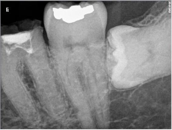
Completely calcified tooth does not necessarily condemn it to extraction

    One must not decide a tooth extraction on the sole basis of an x-ray observation. In cases of complete calcification of root canal systems, the

Category iconD.O.M. versus completely calcified systems,  Dental operating microscope (D.O.M.) assisted R.C.T.,  Extreme endo clinical cases,  Popular Cases Tag iconcalcified,  chelating agent,  Endodontist,  Expertise,  Microscope,  Protaper,  rc prep,  root canal entries,  root canal procedure,  Root canal system calcifications,  root canal therapy,  Root canal treatment,  russian red,  tooth,  zeiss

  • « Go to Previous Page
  • Page 1
  • Page 2
  • Page 3
  • Page 4
  • Page 5
  • Interim pages omitted …
  • Page 22
  • Go to Next Page »

Primary Sidebar

Quality Endodontics

 30+ Years of Experience
in Root Canal Treatment

 300+ Clinical Cases
Published for dentists

 Conveniently Located
Near Metro Vendôme

Call for an appointment:

(514) 667-9541

Cases with high level of difficulty:

Pierre Pizem Endodontics

A little message from Dr Pizem:

« I understand how stressful going to the dentist could be for you, the patient. So let me reassure you. I’ve built my entire reputation on my professionalism and the passion I pour into my craft. I will not compromise on equipment quality, nor on the effort necessary to restore your teeth. In the end, my greatest satisfaction is to see you fully recover your dental health, so you may grab big bites at life again. »

Ready to start your treatment?

In order to avoid risks of further complications, we recommend you book a visit as early as you can. However, if you're already feeling pain or urgent symptoms, we'll make every attempt to see you today.

Get in touch

Footer

Contact

(514) 667-9541

REQUEST APPOINTMENT

Monday
8:30 am - 4:30 pm
Tuesday
8:30 am - 4:30 pm
Wednesday
8:30 am - 4:30 pm
Thursday
8:30 am - 4:30 pm
Friday
Closed
Sat. Sun.
Closed

Clinic info

  • Home
  • About
  • Dr Pizem
  • Publications
  • Referring Dentists
    • Refer a patient
    • Clinical cases
  • Français

Patient info

  • Root Canal Therapy
  • General Dentistry
  • Endodontic Retreatment
  • FAQ New Patients
  • Tooth Saving Tips
5025 Sherbrooke St. W, Suite 330A
Montreal (QC) H4A 1S9
Near Metro Vendôme       Directions

Copyright © 2025 Endomontreal

Return to top