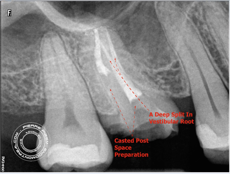
First maxillary molars typically have 4 canal entries and four distinct canals. This molar has only two canal entries and a deep split in its
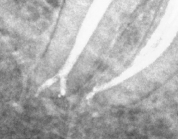
Cleaning, shaping and filling of an apical root canal split
Apical split has been cleaned and shaped with precurved stainless steel Mani endodontic K files. Passive ultrasonic irrigation with NaOCl 5%.
An Endodontic Procedure on a Unique Root Canal Morphology
This case has been posted on a specialized root canal FB forum and earned 596 likes from dentists around the world. See this case at: International
How the Use of Opmi PROergo Dental Operative Microscope Prevented an Unnecessary Molar Extraction. An Endodontist Case Study.
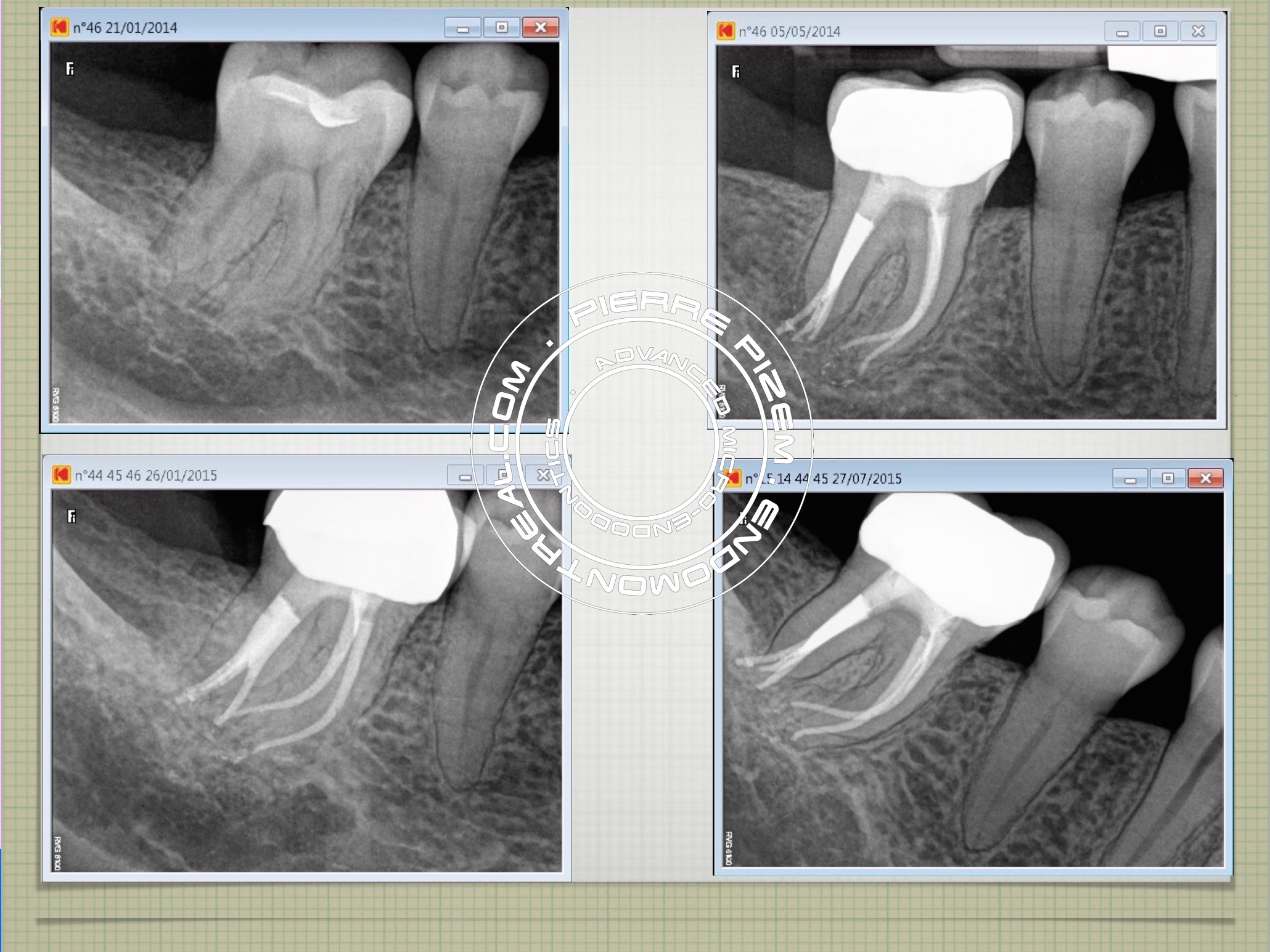
An intricate root canal procedure. Case study number 524236 Two successive root canal procedure attemps failed on this mandibular molar. In
Carl Zeiss OPMI PROergo Insured Enough Visual Accuracy to Prevent a Missed Apical Split in a Calcified Mesial Root.
Endodontist. Case Study Number 197337 The recent addition of dental operative microscope
An Endo Retreatment with Four MTA (Mineral Trioxide Aggregate) Apical Plugs Helped in Preserving this Second Mandibular Molar
Case Study Number 397137 Tooth history: First attempt of RCT on this mandibular second molar in 2005 did not eliminate symptoms, a second attempt
A Vertucci’s Type V Canal Configuration on a Second Maxillary Premolar
Case Study Number 473515 OPMI PROergo from Carl Zeiss allowed us to clearly see the apical split. Each branch has been shaped, cleaned and filled
Carl Zeiss Opmi Proergo Microscope VS Complete Stenosis of an Apical Root Canal Split
Calcified Canals. Case Study Number 487445 Clinical examination: Sinus tract, mobility: 0, deciduous amalgam restoration. Radiographic
Two Distinct Right-Angled Root Canals Exits in First Molar Distal Root (Case 455336)
Case Study Number 455336 Irreversible pulpitis, deep carie, deciduous restoration, broken lingual wall. First molar with 4 root canals. Two mesial


