• Skip to primary navigation
  • Skip to main content
  • Skip to primary sidebar
  • Skip to footer

  (514) 667-9541

  REQUEST APPOINTMENT

HOME

ABOUT

DR PIZEM

FRANÇAIS

Endomontreal Root Canal Clinic logo

ENDOMONTREAL

Dr Pizem Advanced Endodontics Clinic

  • Advanced Root Canal Treatment
    • Symptoms & Procedure
    • Why Micro Endodontics?
  • General Dentistry
  • Patients info
    • Frequently asked questions (FAQ)
    • Need a root canal revision?
    • Treatment sequence at our clinic
    • Tooth saving tips
  • For Dentists
    • Refer a patient
    • Clinical cases
    • Implants vs Endodontics
Contact Us

Gutta percha

You are here: Home / Archives for Gutta percha

August 12, 2011

Endodontic Procedure on a Severely Curved Distovestibular Root Canal of a Calcified Maxillary Molar

Root Canal Procedure. Case Study Number 492626 Patient is having an AAA with a necrotic pulp, an extensive deciduous restoration and a huge tooh

Category iconCurved canals,  D.O.M. versus partially calcified systems,  Dental operating microscope (D.O.M.) assisted R.C.T.,  Extreme endo clinical cases,  Extremely curved root canals (90 degrees +),  NEW CASES,  Popular Cases,  Striving for second Mesio vestibular (MB2) Tag iconaaa,  because,  Calcium hydroxide,  Carl Zeiss,  curvatures of greater than 90 degrees,  Dental expertise,  Dental operative microscope,  Dystrophic calcifications,  Endodontic expertise,  exudation,  franklin s weine,  Gutta percha,  Mb2,  Mesio vestibular,  Mesiovestibular,  microendodontics,  opmi proergo,  private dental insurance,  pro taper,  Protaper dentsply,  pulp canal sealer,  root canal procedure,  root canal sealer,  Root canal system calcifications,  Root canal treatment,  schneider sw,  Second mesio vestibular

March 22, 2011

Carl Zeiss Opmi Proergo Microscope VS Complete Stenosis of an Apical Root Canal Split

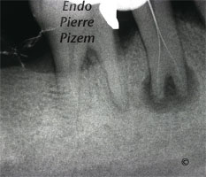
Calcified Canals. Case Study Number 487445 Clinical examination: Sinus tract, mobility: 0, deciduous amalgam restoration. Radiographic

Category iconAtypical canal configurations,  D.O.M. versus completely calcified systems,  Dental operating microscope (D.O.M.) assisted R.C.T.,  Extreme endo clinical cases,  NEW CASES,  Taurodontism,  Type V Tag iconabsorbent paper points,  apical delta,  bull tooth,  calcified canals,  Calcium hydroxide,  complete root canal stenosis,  Dental expertise,  Dental operative microscope,  dental radiograph,  Dystrophic calcifications,  Endodontic expertise,  endodontic file,  Endodontics,  Gutta percha,  hypercementosis,  hypertaurodontism,  image,  microendodontics,  Pain,  periapical radiograph,  radiolucent apical lesion,  root canal apical split,  root canal procedure,  Root canal system calcifications,  Sinus tract,  vertucci type V root canal configuration

December 15, 2010

Mesial Calcified Canals Entries Location Under High Magnification

  An intricate root canal procedure, because this pre operative condition involves dealing with complete canal stenosis caused by dystrophic

Category iconD.O.M. versus completely calcified systems,  Dental operating microscope (D.O.M.) assisted R.C.T.,  NEW CASES Tag iconbecause,  calcified canals,  chelator,  complete root canal stenosis,  Dental operative microscope,  Dystrophic calcifications,  edta,  endo buchanan,  Endodontic expertise,  Endodontics,  Gutta percha,  Images,  Location,  maillefer,  microendodontics,  pro taper,  pulp canal sealer,  rc prep,  root canal procedure,  Root canal system calcifications

July 21, 2008

Dealing with a Crowned Maxillary Molar and Extreme Calcifications in it’s Root Canal System

Endodontic Procedure Under Dental Operative Microscope. Case Study Number 282816 Surgical operating microscope assisted root canal treatment through

Category iconD.O.M. versus completely calcified systems,  Dental operating microscope (D.O.M.) assisted R.C.T.,  NEW CASES Tag iconaccess difficulties,  Coronal-radicular access,  Dental operative microscope,  Dystrophic calcifications,  endodontic files,  endodontic procedure,  Gutta percha,  maxillary molar,  Opmi Proergo microscope,  surgical operative microscope

July 7, 2008

An Endodontic Revision and a Tooth Sectioning on a Mandibular Right First Molar to Preserve it

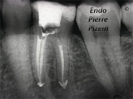
Endodontic Re-treatment and Tooth Sectioning Procedure (2004). Case Study Number 336 Root canal retreatment in distal root, machined post

Category iconDental operative microscope and retreatment,  Post endodontic treatment outcomes,  Regeneration of the periradicular tissues,  Tooth sectioning Tag iconendodontic retreatment,  endodontic revision,  Endodontist,  Gutta percha,  Images,  microendodontics,  Retreatment,  root canal procedure,  surgical endodontics,  tooth sectioning

June 29, 2008

Fiberglass Reinforced Composite Posts Removal in Both Maxillary Premolar to Allow Endodontic Retreatment

Root Canal Procedure. Case Study Number 1 First premolar was having an under fill in its distovestibular root canal. Meaning that one small diameter

Category iconAtypical canal configurations,  D.O.M. versus completely calcified systems,  Dealing with casted and machined posts removal,  Dealing with ledges and apical zip,  Dental operating microscope (D.O.M.) assisted R.C.T.,  Dental operative microscope and retreatment,  Extreme endo clinical cases,  Regeneration of the periradicular tissues Tag iconaaa,  Dental operative microscope,  Dystrophic calcifications,  endodontic retreatment,  endodontic revision,  fiberglass reinforced composite post,  Gutta percha,  Location,  post removal,  premolar,  pulp canal sealer,  root canal procedure,  underfill

June 14, 2008

Lower Left Second Molar Vertucci’s Type V Canal Configuration

Intricate Endodontic Procedure. Case Study Number 417 A Vertucci type V pulp space configuration can be described as follow: One canal leaves the

Category iconAtypical canal configurations,  Curved canals,  Extreme endo clinical cases,  Extremely curved root canals (90 degrees +),  NEW CASES,  Type II,  Type V,  Very long teeth Tag iconapical delta,  Dental expertise,  endodontic procedure,  Endodontics,  Endodontist,  Gutta percha,  Images,  pulp canal sealer,  root canal apical split,  severe curvature,  vertucci type V root canal configuration,  very long tooth

  • « Go to Previous Page
  • Page 1
  • Page 2
  • Page 3

Primary Sidebar

Quality Endodontics

 30+ Years of Experience
in Root Canal Treatment

 300+ Clinical Cases
Published for dentists

 Conveniently Located
Near Metro Vendôme

Call for an appointment:

(514) 667-9541

Cases with high level of difficulty:

Pierre Pizem Endodontics

A little message from Dr Pizem:

« I understand how stressful going to the dentist could be for you, the patient. So let me reassure you. I’ve built my entire reputation on my professionalism and the passion I pour into my craft. I will not compromise on equipment quality, nor on the effort necessary to restore your teeth. In the end, my greatest satisfaction is to see you fully recover your dental health, so you may grab big bites at life again. »

Ready to start your treatment?

In order to avoid risks of further complications, we recommend you book a visit as early as you can. However, if you're already feeling pain or urgent symptoms, we'll make every attempt to see you today.

Get in touch

Footer

Contact

(514) 667-9541

REQUEST APPOINTMENT

Monday
8:30 am - 4:30 pm
Tuesday
8:30 am - 4:30 pm
Wednesday
8:30 am - 4:30 pm
Thursday
8:30 am - 4:30 pm
Friday
Closed
Sat. Sun.
Closed

Clinic info

  • Home
  • About
  • Dr Pizem
  • Publications
  • Referring Dentists
    • Refer a patient
    • Clinical cases
  • Français

Patient info

  • Root Canal Therapy
  • General Dentistry
  • Endodontic Retreatment
  • FAQ New Patients
  • Tooth Saving Tips
5025 Sherbrooke St. W, Suite 330A
Montreal (QC) H4A 1S9
Near Metro Vendôme       Directions

Copyright © 2025 Endomontreal

Return to top