Endodontist. Case Study Number 474446 Deep deciduous restorations have been replaced 4 days ago. Patient has been experiencing
When a “J” Type Lesion on an X Ray Image, as well as Probing a “Deep Narrow Periodontal Pocket” Could Have Missled the Dental Practioner to Conclude the Presence of a Cracked Tooth.
This is a case where, based on the X ray image of a "J" type lesion in combination with a deep narrow periodontal probing, one could
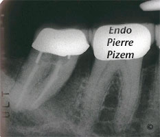
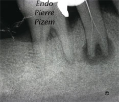
A Huge Radiographic “J” Shape Lesion, Does the Tooth Has a Vertical Root Fracture? a Periodontal Infection? an Endodontic Infection?
Case Study Number 456147 We have been presented with this mandibular second molar that has a periapical lucency which also has a periodontal
Carl Zeiss Opmi Proergo Microscope VS Complete Stenosis of an Apical Root Canal Split
Calcified Canals. Case Study Number 487445 Clinical examination: Sinus tract, mobility: 0, deciduous amalgam restoration. Radiographic

Zeiss Opmi Pro Ergo Dental Operative Microscope VS Almost Complete Stenosis of Root Canal System
Root Canal Procedure on Calcified Canals. Case Study Number 317736 Patient referred for pre prosthetic endodontic treatment on mandibular first
Endodontic Revision on First Mandibular Molar
Case study number 485946 Symptomatic mandibular molar, patient can't chew on that side. Referred to us for endodontic revision. First appointment
Dealing with Dystrophic Calcification in Mesiovestibular Root Canals
Case Study Number 486726 Patient was referred to us with a partial pulpectomy in 3 out of four canals. The case came with a
A very very long tooth. 28 mm in palatal, 26 mm in mesio vestibulars 1 and 2
A gigantic 28 mm treated maxillary molar on display with four mineralized canals incuding the MB2. Its mesio vestibular root has two
The Dental Operative Microscope and a MTA Tooth Perforation Repair. The Impossible Made Possible.
Case Study Number 461716 Patient came to our office with spontaneous intermittent pain on maxillary right side. He can readily identify the tooth,