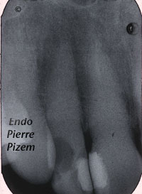
A new Dental Operative Microscope (D.O.M.) Assisted Root Canal Treatment in a Calcified Maxillary Lateral
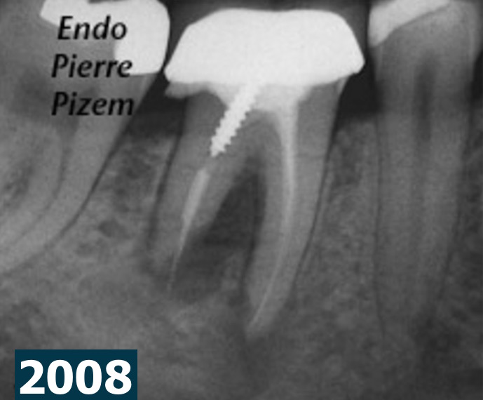
Root Canal Treatment on a Mandibular First Molar Presenting with a Dilacerated Root Canal System
Endodontic. Case Study Number 498946.
Endodontic Therapy on Maxillary Second Premolar With One Canal Dividing in Apical Third. The Infamous Apical Delta
Endodontist. Case Study of Root Canal Procedure Number 362515 Pecora al. in 1993 reported
Microendodontics with Zeiss OPMI PRO Ergo Operative Microscope: Striving to Find a Pathway to the Apices and to Clean a “C” Shape RC system.
The name comes from the letter "C" shape appearance of a very large isthmus in the pulp chamber floor when viewed from above. This isthmus or groove
An Intricate Root Canal Procedure on a Mineralized Second Maxillary Molar with a Canal Curvature into an “S” Form
Endodontic Procedure. Case Study Number 449927 To treat such a tooth in endodontics we needed to deal with: Difficult access Long tooth
An Intricate Root Canal Procedure on a Severely Curved Root Canal System with Pulp Tissue Fibrosis
MicroEndodontic. Case Study Number 500047 Canal curvatures are a challenge to preparation and can be the origin of many technical complications
Microendodontics with Carl Zeiss OPMI PROergo Dental Operative Microscope. Root Canal Treatment Procedure on a Tooth with Calcified Canal System on Pre-Op. Radiograph
A new Dental Operative Microscope (D.O.M.) assisted root canal treatment in a calcified maxillary
Good Prognosis is in the eye of the beholder
Case Study Number 430646 External root resorption associated with chronic apical periodontitis altered the shape and position
Microendodontics with CARL ZEISS OPMI PROergo VS Completely Calcified Canal
A New Dental Operative Microscope (D.O.M.) Assisted Root Canal Treatment on Maxillary Incisor with a Calcified Canal. MicroEndodontic. Case