Thanks for visiting this blog on endodontics We wish you all a happy new year!
Radix Entomolaris, Calcified Canals and the Usefulness of a Dental Operative Microscope (D.O.M.)
Endodontic. Case Study Number 505146 A peculiar anatomical variation can be noticed on this mandibular first molar. What appeared to look like
Sven-Erick Hamp Class III Furcation Defect? Parodontal Prognosis? A Seven years Follow Up
Preoperative X ray dental film shows a "furcation defect" encompassing the entire width of the tooth (no probing). A root canal treatment implying a
To Save or Not To Save? That Was the Question. A Seven Years Post Endodontic Treatment Outcome Follow Up
Case Study Number 368745 Patient was told seven years ago to remove lower right premolar and replace this tooth by an implant supported
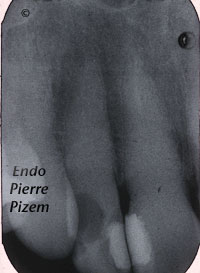
Microendodontics with Carl Zeiss OPMI PROergo Dental Operative Microscope. Root Canal Treatment Procedure on a Lateral Incisor with a Calcified Canal
A new Dental Operative Microscope (D.O.M.) Assisted Root Canal Treatment in a Calcified Maxillary Lateral
Root Canal Treatment on a Mandibular First Molar Presenting with a Dilacerated Root Canal System
Endodontic. Case Study Number 498946.
Endodontic Therapy on Maxillary Second Premolar With One Canal Dividing in Apical Third. The Infamous Apical Delta
Endodontist. Case Study of Root Canal Procedure Number 362515 Pecora al. in 1993 reported
Microendodontics with Zeiss OPMI PRO Ergo Operative Microscope: Striving to Find a Pathway to the Apices and to Clean a “C” Shape RC system.
The name comes from the letter "C" shape appearance of a very large isthmus in the pulp chamber floor when viewed from above. This isthmus or groove
Intricate Root Canal Procedure on Root Canals Curvatures with Very Small Radius.
Endodontist. Root Canal Procedure. Case Study Number 49821617 Note on the post operative Xray dental film, the dilacerated apical curves in